-
2025-09-10 Sept
Light-Powered Tools for Spatial Omics
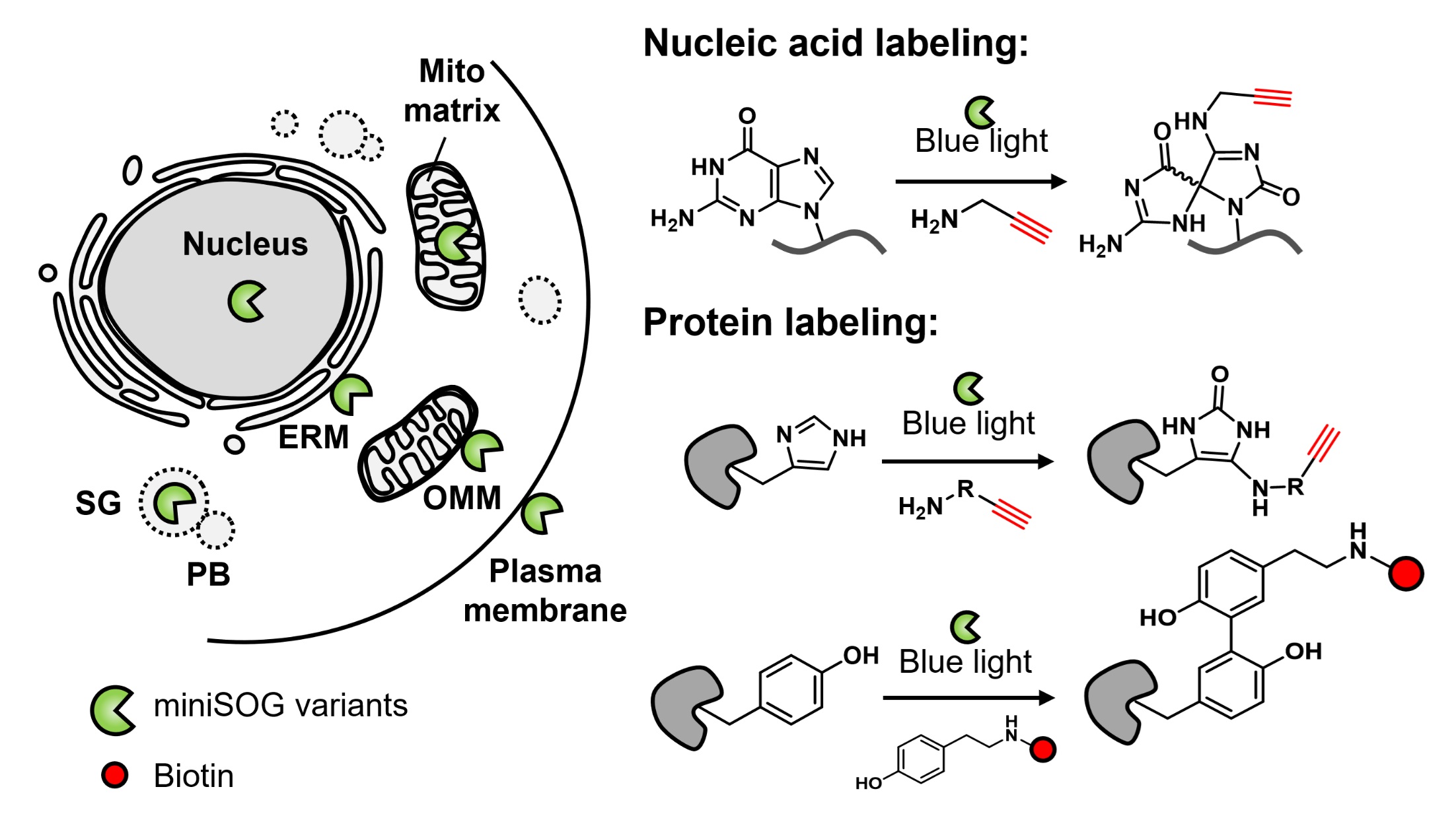
Light offers powerful opportunities for mapping biomolecules in living systems with high spatial and temporal precision. In a recent Accounts of Chemical Research publication, we review the development of genetically encoded photocatalytic proximity labeling (PPL) methods that use light or bioluminescence to map RNAs, proteins, and multi-omic landscapes in cells and animals. Key advances include CAP-seq for transcriptome profiling, RinID for proteomic mapping, and the luminescence-activated LAP platform for in vivo studies. These tools enable rapid, pulse-chase labeling with second-scale resolution, revealing localized mRNA translation, protein turnover, and cell-cell interactions in living systems. Looking ahead, efforts will focus on engineering red-shifted photosensitizers for deep-tissue imaging, split-enzyme designs for studying organelle contact sites, and orthogonal labeling chemistries for multiplexed spatial omics.
-
2025-08-15 Aug
Bright New Tools for Voltage Imaging
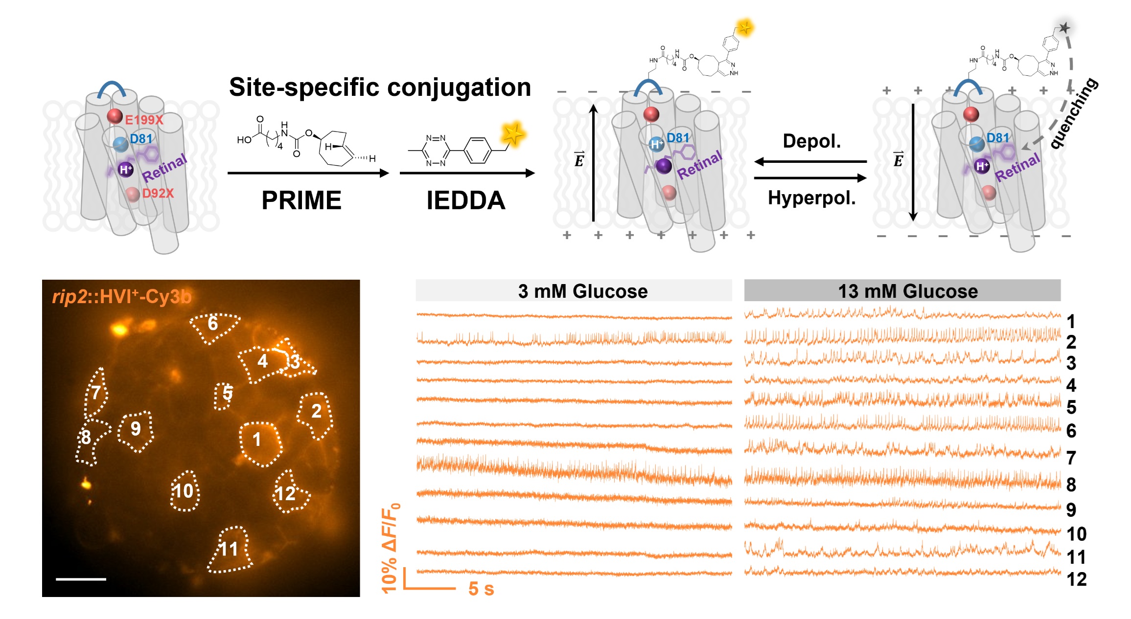
In a recent Science Advances publication, we report HVI+, a new class of voltage indicators that light up when cells fire. Unlike traditional indicators that dim upon depolarization, HVI+ grows brighter, reducing background noise and enabling clearer recordings of fast electrical signals. The lead variant, HVI+-Cy3b, achieves 55% ΔF/F0 per 100 mV, the highest sensitivity among positive-going indicators. HVI+ supports ratiometric imaging in beating cardiomyocytes for motion-corrected action potential recordings and, when paired with negative-going indicators, reveals cell-type-specific voltage responses in pancreatic islets under glucose stimulation. These advances provide powerful tools for mapping electrical activity in excitable cells with high precision.
-
2025-07-03 July
Red Light Powers Next-Generation Protein Mapping
Red light penetrates biological tissues far deeper than blue light—and we have now harnessed this property to map proteins inside living cells with high spatial resolution. In a recent Analytical Chemistry study, we introduced FLAPP, a technique that uses near-infrared light to trigger fast, proximity-based labeling of proteins exactly where they are in the cell. Unlike conventional blue-light methods that struggle with background signals and limited tissue reach, FLAPP enables us to profile hundreds of proteins in the nucleus and mitochondria with 96% spatial specificity, even through millimeter-thick tissue slices. By combining deep-tissue red light activation with genetic targeting, we provide a powerful new tool for studying protein networks in complex biological environments.
-
2025-06-26 June
Congratulations, Class of 2025!
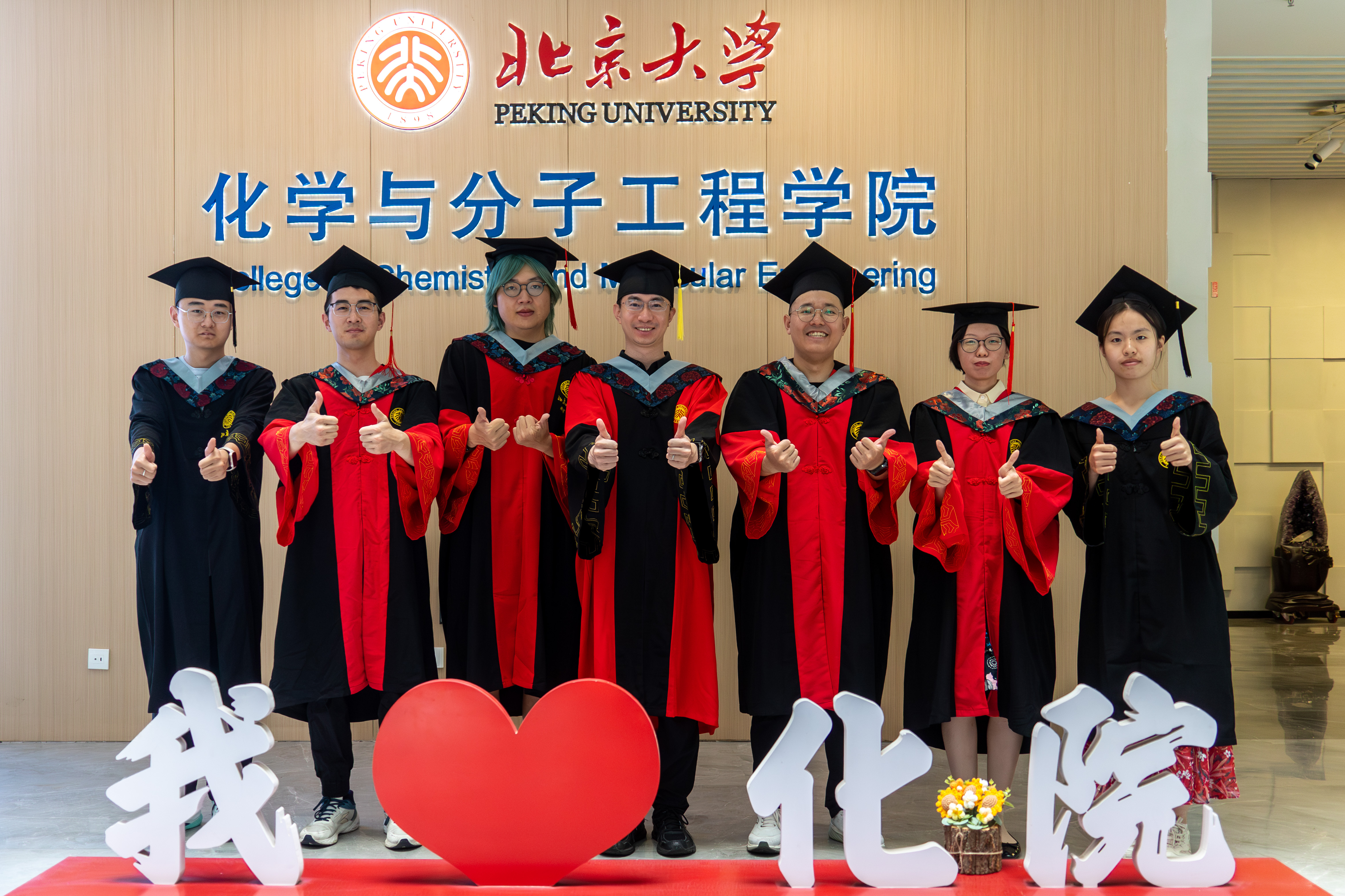
We are delighted to celebrate the achievements of our lab members this year! Drs. Yu Chen, Chen Li, Fu Zheng, and Siyan Zhu successfully defended their doctoral theses, marking an important milestone in their academic journeys. In addition, undergraduate students Ruixin Zhu and Xinhao Li completed their Bachelor of Science degrees at PKU and will continue their studies at CCME as Ph.D. candidates. We are proud of all of you and look forward to your future successes!
-
2025-05-26 May
Lighting Up Cell’s Hidden Architecture with Bioluminescence

A detailed mechanistic picture of proteins, RNAs, and cells hinges on accurately mapping their spatial organization and interactions. Although photocatalytic proximity labeling (PL) offers spatiotemporally controlled tagging of biomolecules and cells, its reliance on external light limits deep-tissue applications in live animals. To address this, we harnessed luciferase-generated bioluminescence as an internal light source, obviating the need for external illumination. In a study recently accepted at the journal Chem, we introduce a biocompatible, bioluminescence resonance energy transfer (BRET)-driven PL platform that enables spatially selective tagging at both the intracellular and intercellular levels in vitro and in vivo. By combining the high resolution of photocatalytic labeling with the physiological compatibility of luciferases, this toolkit paves the way for deep-tissue, high-resolution spatial omics in living systems.
-
2024-06-30 June
Eight Graduates, Countless Discoveries Ahead!
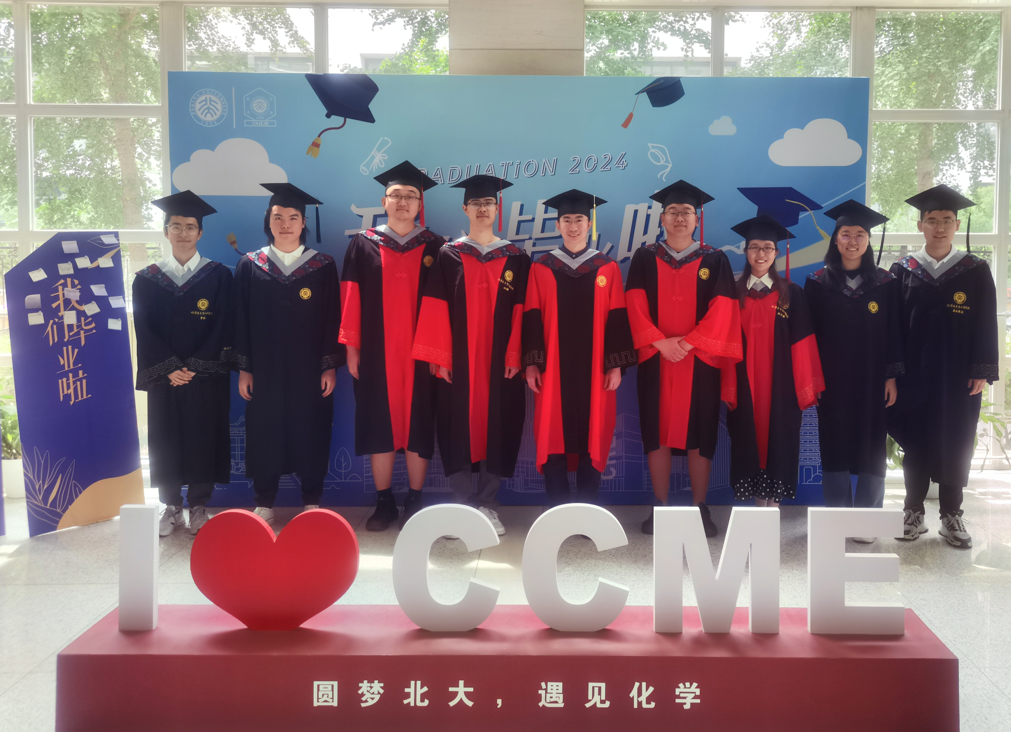
This year brought an exciting chapter for our lab as eight members graduated and set off on new paths. Four PhD students—Chang Lin, Ziqi Ren, Xinyue Zhou, and Ruixiang Wang—completed their doctoral studies, while undergraduates Chang Cao, Haoyu Yang, Keyuan Ren, and Jinsaibo Gong earned their bachelor’s degrees. Chang Lin will soon join Stanford University as a postdoctoral researcher, and Keyuan is headed to the University of Chicago for her PhD. Staying closer to home, Xinyue, Ruixiang, Chang Cao, Haoyu, and Jinsaibo will continue their academic journeys here at CCME, diving into new projects and collaborations. We’re excited to see the discoveries they will bring to life in the years ahead.
-
2023-12-12 Dec
Moving into New Lab Building!
Our lab has moved to the new research laboratory building at CCME! The new space is divided into four areas: the Biochem Lab and Chem Biol Lab are on the fifth floor, next to our offices, while the tissue culture room and imaging room are located in the basement. Despite the substantial effort required to move equipment and reagents, we managed to settle down within a week. Thank you everyone for doing a fantastic job!
-
2023-12-11 Dec
A Protein Map for the Axon Initial Segment

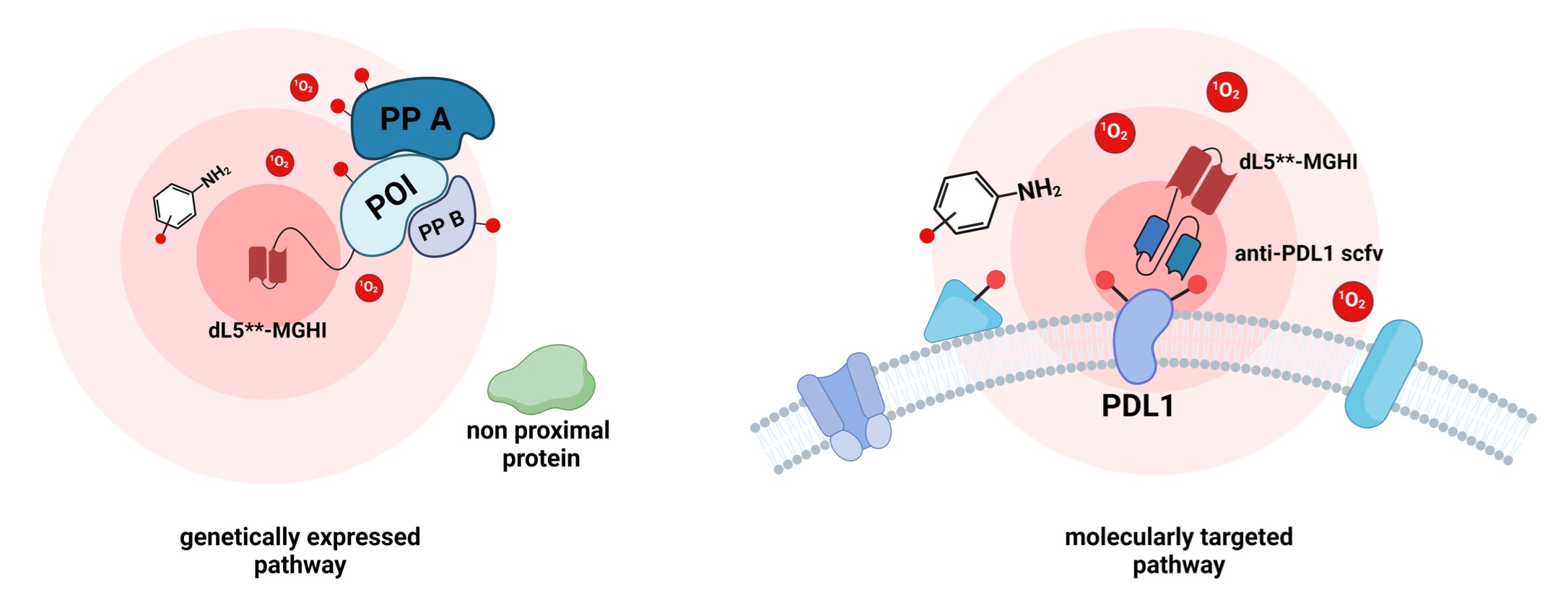
In the past, we have used voltage imaging to visualize the initiation of action potentials at the axon initial segment (AIS). Recently, we have shifted our focus to investigating the molecular inventory of this intriguing subcellular compartment. In a publication in Nature Communications, we introduced an immunoproximity labeling approach that targets the endogenous AIS protein neurofascin for precise and high-resolution mapping of AIS proteins. Immuno-PL has enabled us to uncover AIS protein components and their dynamic changes throughout neuronal development. The instructional proteome has empowered us to discern authentic AIS-enriched proteins, including SCRIB, a discovery validated through collaboration with Professor Matthew Rasband’s research group at Baylor College of Medicine. This powerful and adaptable approach not only refines our understanding of the AIS proteome but also serves as a valuable resource to unravel the intricate mechanisms that regulate the structure and function of the AIS.
-
2023-11-22 Nov
Red Indicators for Voltage Imaging in Tissues
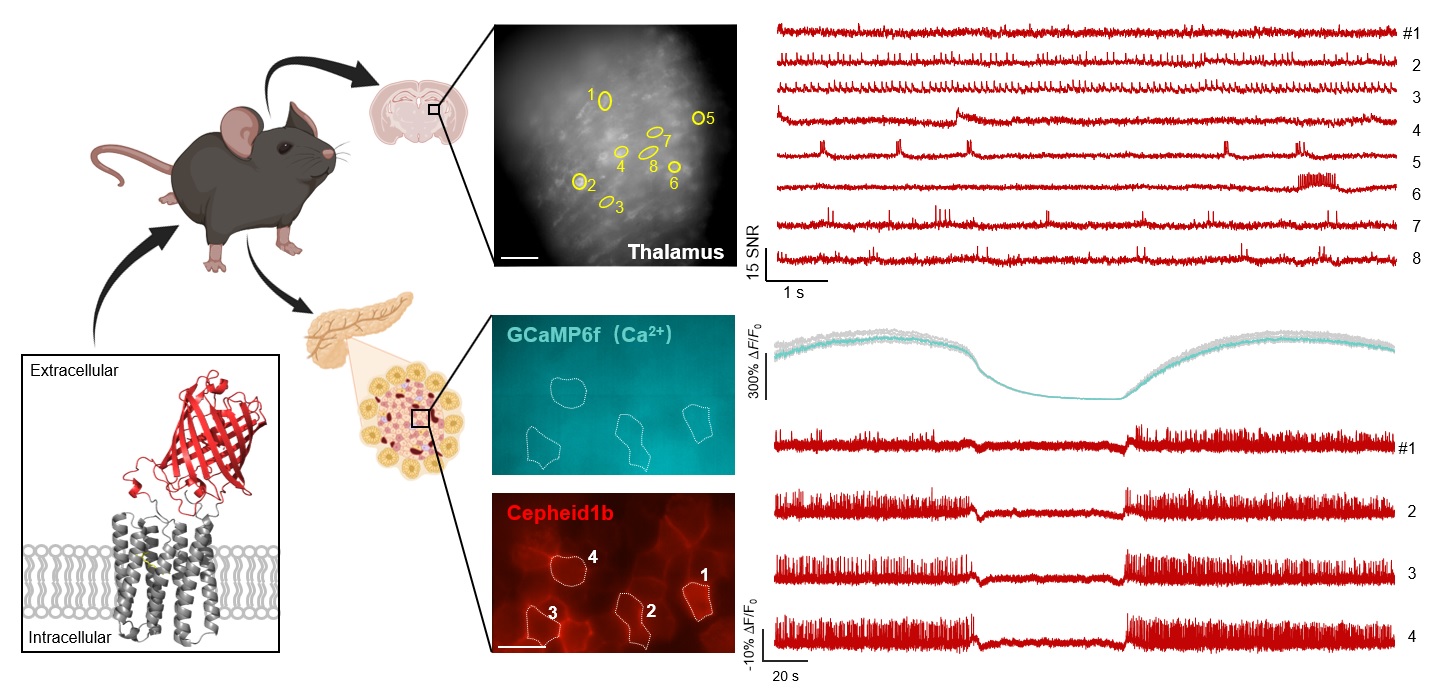
To enhance the voltage sensitivity of eFRET GEVIs, we explored a novel approach involving the insertion of red fluorescent proteins into the extracellular loop of rhodopsin proteins. This unconventional method yielded a surprisingly high dynamic range compared to the conventional practice of appending fluorescent proteins to the termini. In the recent issue of Science Advances, we present the engineering of these red eFRET GEVIs, named Cepheid1b and Cepheid1s, which exhibit improved voltage response, brightness, and photo-stability. These advancements facilitate voltage imaging in mouse brain slices with minimal laser power, uncovering burst firing and subthreshold depolarization activities in multiple neurons. Notably, Cepheid indicators support multiplexed imaging alongside green fluorescent indicators for calcium or glutamate, offering comprehensive insights into cellular dynamics. Furthermore, their compatibility with CheRiff, a spectrally orthogonal optogenetic actuator, enables all-optical electrophysiology measurements of neuronal excitability. In a notable application, Cepheid1b enables non-invasive observation of glucose-stimulated correlations between electrical spiking and calcium oscillations in pancreatic islet tissue. These findings mark a significant advancement in voltage imaging technology, promising deeper insights into cellular function and opening avenues for innovative neuroscientific and biomedical research endeavors.
-
2023-11-15 Nov
Mapping RNAs in Stressed Cells
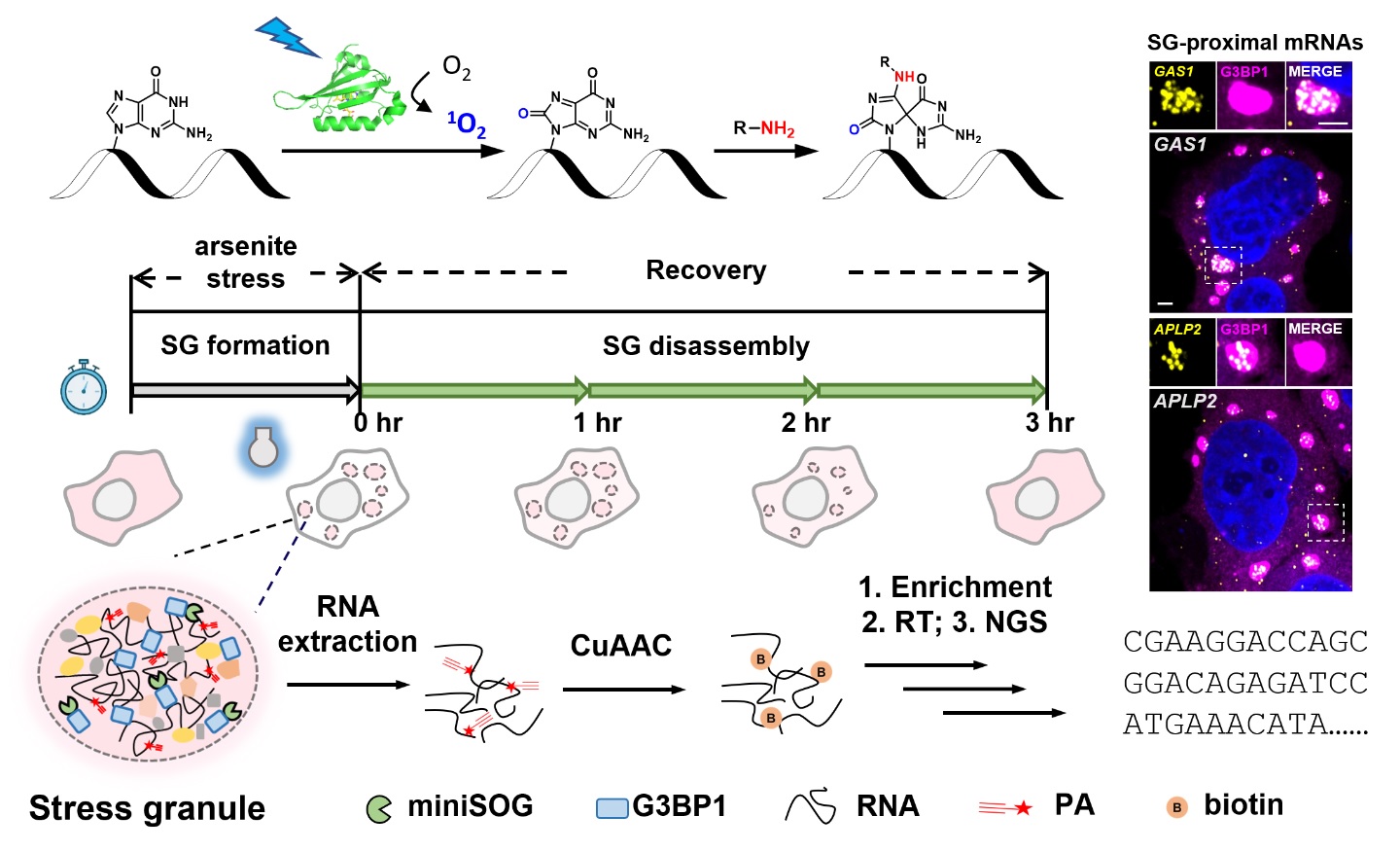
When cells encounter stressors such as oxidation, osmotic shock, or starvation, they activate a series of response pathways, including the formation of stress granules (SGs). These SGs are membraneless organelles vital for cellular functions and are often implicated in neurodegenerative diseases. Understanding the molecular composition of SGs is essential for comprehending their biological role in stress response. In a recent publication in Nature Communications, we utilized the photocatalytic RNA labeling and sequencing method, CAP-seq, to systematically profile the transcriptome of SGs in live cells with unprecedented spatial resolution. CAP-seq analysis revealed stress-specific mRNA targeting to SGs under conditions such as oxidative stress or osmotic shock. Furthermore, CAP-seq unveiled the temporal dynamics of RNA localization during SG disassembly, revealing a model of AU-rich, translationally repressed SG nanostructures persisting after stress cessation. These findings shed new light on the molecular mechanisms underlying SG function in cellular stress responses and neurodegenerative disorders.
-
2023-11-08 Nov
Adding Spatial Dimensions to Protein Turnover
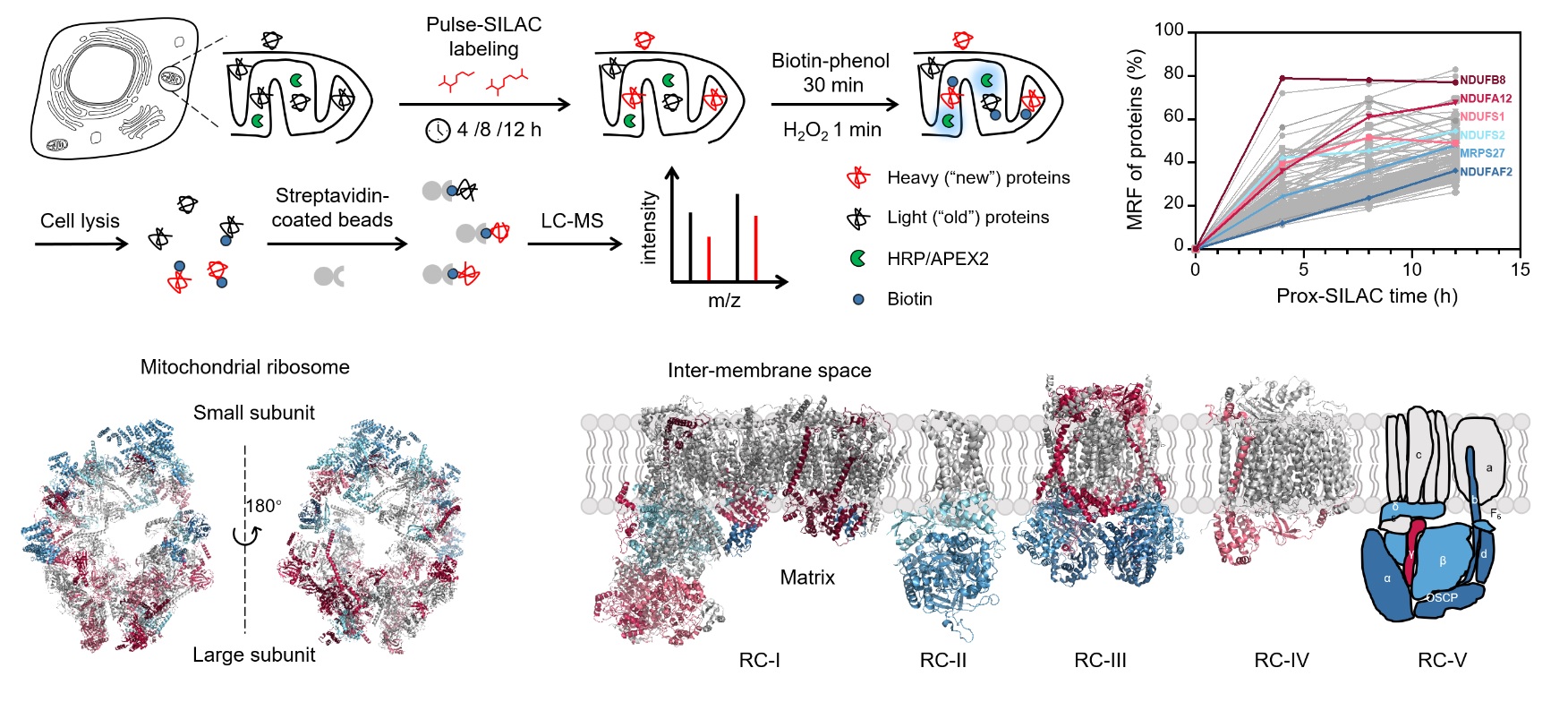
Eukaryotic cells, with their highly compartmentalized nature, rely on precisely regulated protein trafficking for their proper functioning. As a prime example, the secretory pathway orchestrates the journey of membrane receptors from synthesis in the endoplasmic reticulum (ER) to their eventual destination at the plasma membrane, involving crucial stops at the Golgi apparatus and vesicles. While existing chemical tools have enabled the measurement of protein turnover at the whole-cell level, they often lack the much-needed subcellular spatial resolution. In the recent issue of Nature Communications, we describe the development of prox-SILAC technique, a fusion of proximity labeling and pulse-SILAC metabolic tagging, which promises to fill this gap. Prox-SILAC reveals the intricate dynamics of protein turnover within cellular organelles, including the mitochondria and the ER, shedding new light on cellular activities under both physiological and pathological conditions.
-
2023-08-27 Aug
Super-Temporal Resolution Voltage Imaging
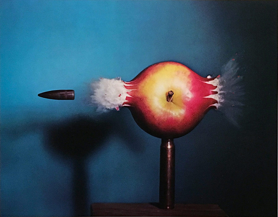
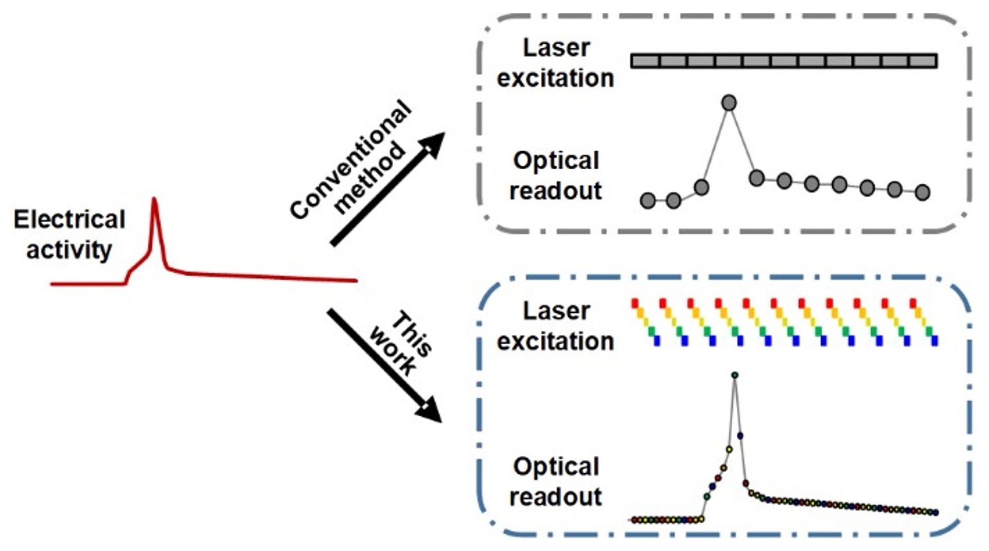
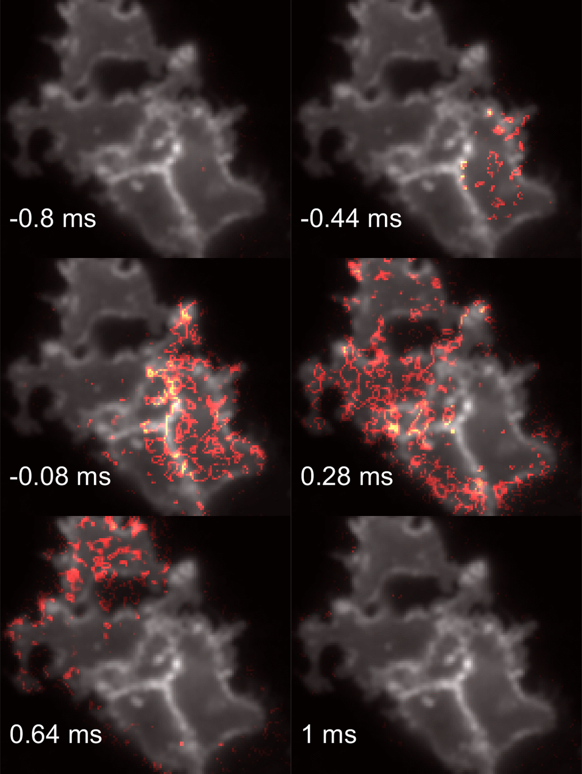
Spatiotemporally resolved mapping of neuronal action potentials (APs) not only requires highly sensitive voltage indicators, but also depends on high-speed imaging apparatus. Ideally, the acquisition rate of voltage imaging should exceed 1 kilohertz to capture the millisecond timescale APs. However, even with the fastest cameras that money can buy, such as scientific CMOS cameras, the full frame acquisition rate is often limited to 100 Hz. While higher speed could be achieved by reducing the number of pixels, this comes with the price of smaller field of view. To address this challenge, we came up with a strategy to boost the effective camera acquisition rate via stroboscopic illumination scheme. Our idea was inspired by high-speed stroboscopic photography, where the object is briefly illuminated with a flash of light, whose duration is much shorter than the camera shutter. The temporal precision is thus defined by the pulse duration of flashed light rather than the camera shutter. This technique is most famously shown by the photo Bullet through Apple, made by Harold E. Edgerton back in 1964. Published in the recent issue of Chem. Biomed. Imaging, our stroboscopic voltage imaging technique achieved 5-fold improvement in the temporal resolution, allowing the mapping of both intercellular and intracellular electrical signaling. This paper has been selected as ACS Editor's Choice.
-
2023-08-20 Aug
More Stable Hybrid Voltage Indicators
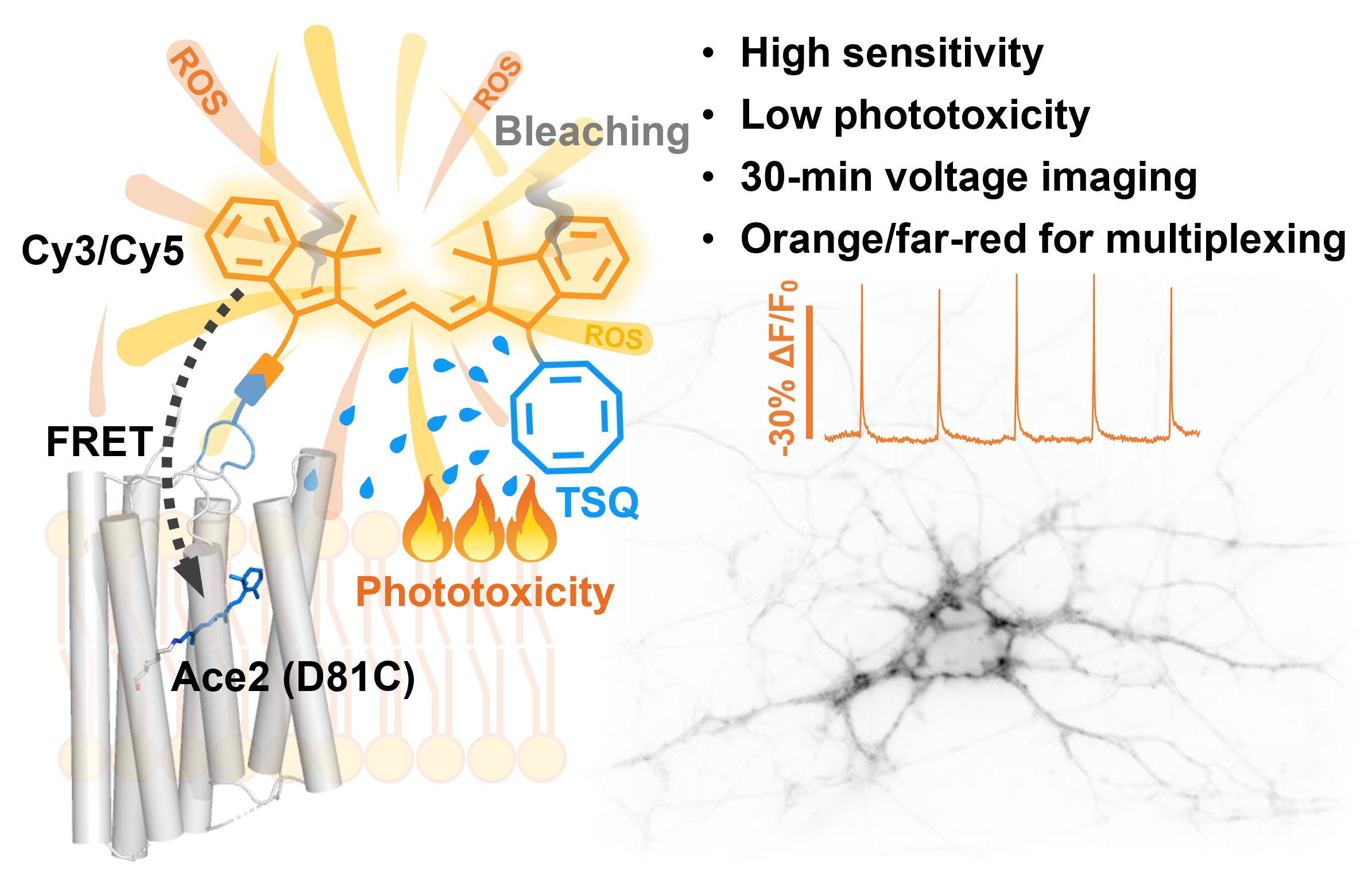
Voltage imaging often operates in shot noise-limited regime with a tight photon budget. Hybrid voltage indicator (HVI) design aims to solve this problem by employing bright and photostable synthetic fluorophores. In the recent issue of PNAS, we report upgraded versions of HVIs that enable continuous voltage imaging for 30 min. This work is in collaboration with Prof. Zhixing Chen's group, who developed cyclooctatetraene-conjugated dyes with substantially improved photostability and reduced phototoxicity.
-
2023-07-27 July
Imaging-Guided Cell Selection
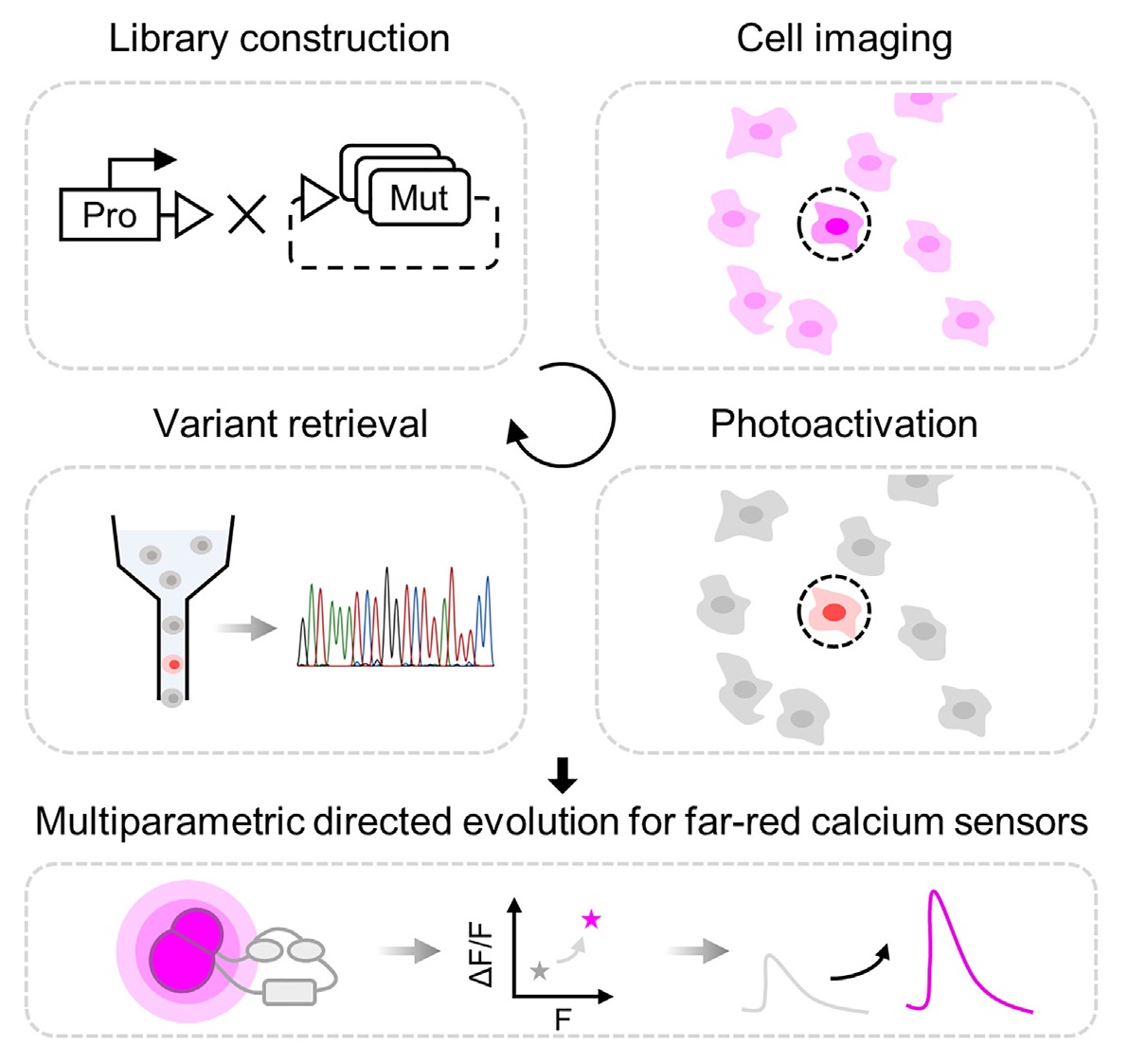
Seeing is believing. Our understanding of cellular physiology has been greatly advanced with the development of optical imaging instruments. It is now possible to detect small variations in functional phenotypes within a heterogeneous cell population, which could arise from alterations at the genetic, epigenetic or transcriptomic levels. However, it remains challenging to efficiently link the observed phenotype to the underlying genotype. To address this challenge, we have built a microscope to allow imaging-guided cell selection. Published in Cell Reports Methods, we describe the development and applications of such method in the context of engineering genetically encoded indicators.
-
2023-06-29 June
Congratulations to Our Graduates!

We have a record-high number of graduates this year! Congratulations to Shuzhang, Gang, Junqi, Luxin, Ruixuan, and Yi for defending their PhD thesis. Thank you all for your hard work and contributions to the lab over the past years. Congratulations to Han, Peiyuan, Ruijie, and Wenyu for defending their undergraduate thesis! All the undergraduates are enrolling as first-year graduate students at CCME this coming September.
-
2023-06-18 June
Peng Receives CCS Life Chemistry Award
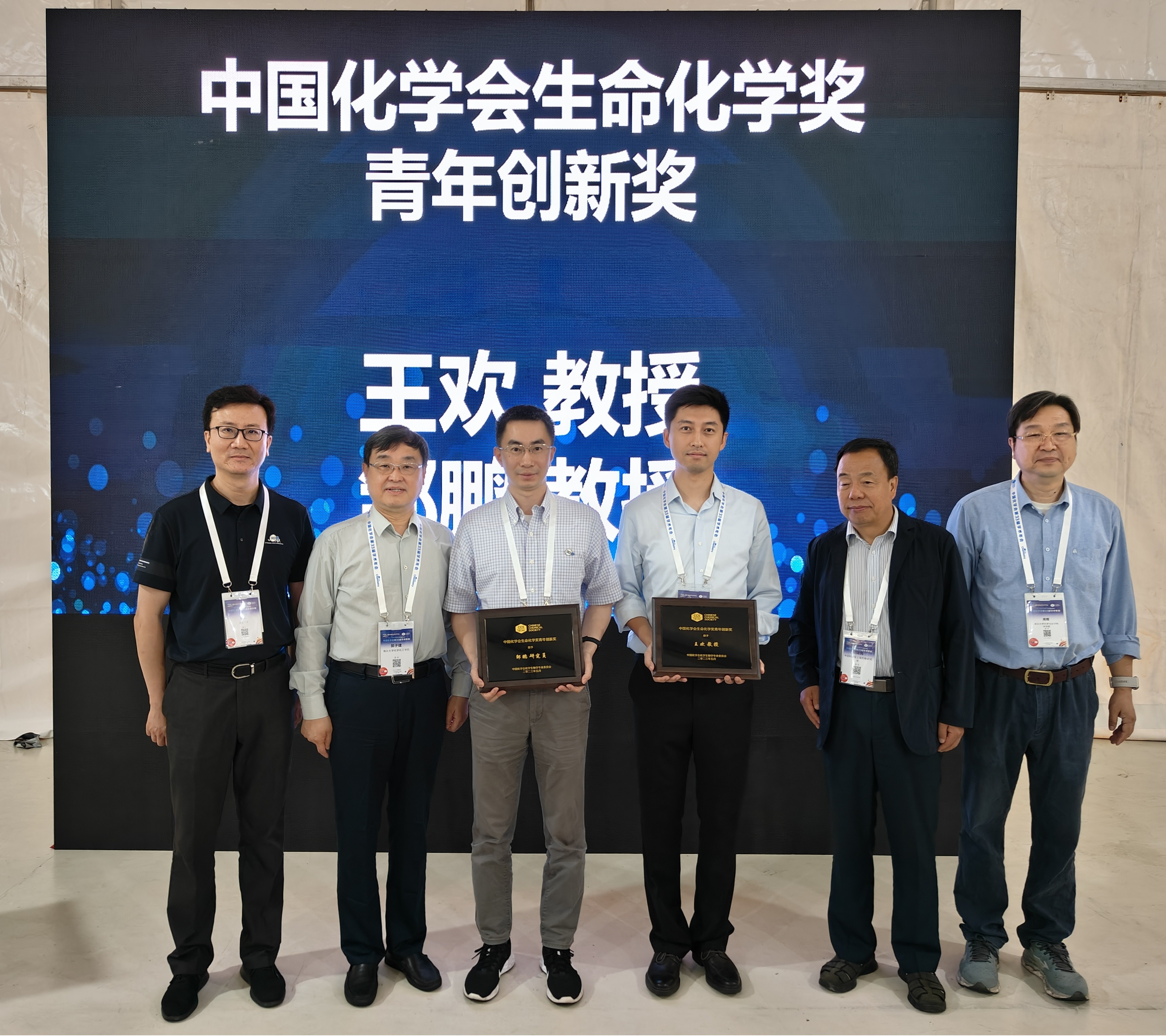
Peng is humbled and honored to receive the Life Chemistry Award from Chinese Chemical Society. The award is presented by the Chemical Biology Committee during the 33rd Chinese Chemical Society Congress in Qingdao. We are both deeply grateful and immensely encouraged by the recognition of our work on chemical neuroscience.
-
2023-05-30 May
RNA in the ER
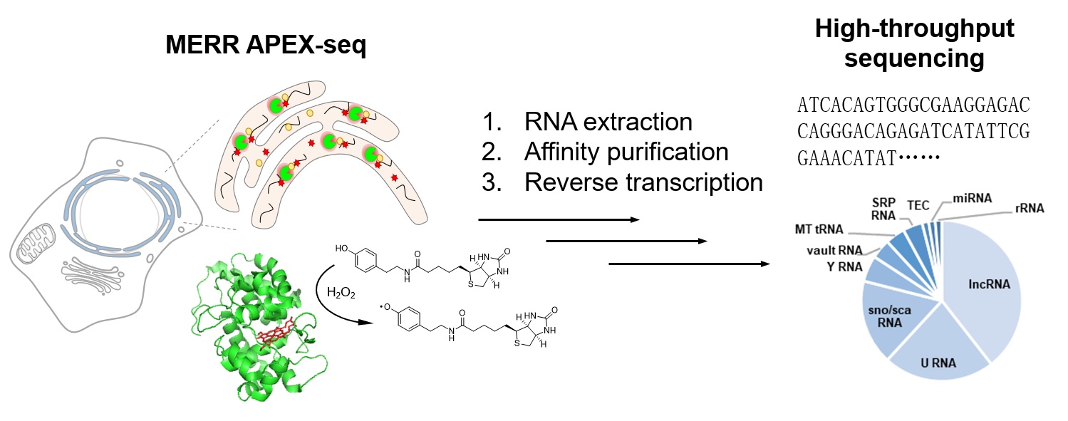
Despite broad distributions throughout the cytoplasm, RNA molecules are conventionally believed to be excluded from the secretory pathway compartments including the endoplasmic reticulum (ER). Recent discovery of RNA N-glycan modification (glycoRNAs) has challenged this view, but direct evidence of RNA localization in the ER lumen has been lacking. To resolve this issue, we applied our recently upgraded version of MERR APEX-seq with enhanced sensitivity to the ER. Published in Biochemistry, we were surprised to identify small non-coding RNAs in the ER lumen of human embryonic kidney 293T cells and rat cortical neurons. These include U RNAs and Y RNAs, which raises interesting questions regarding their transport mechanism and biological functions in the secretory pathway.
-
2023-05-23 May
Photocatalytic Protein Labeling
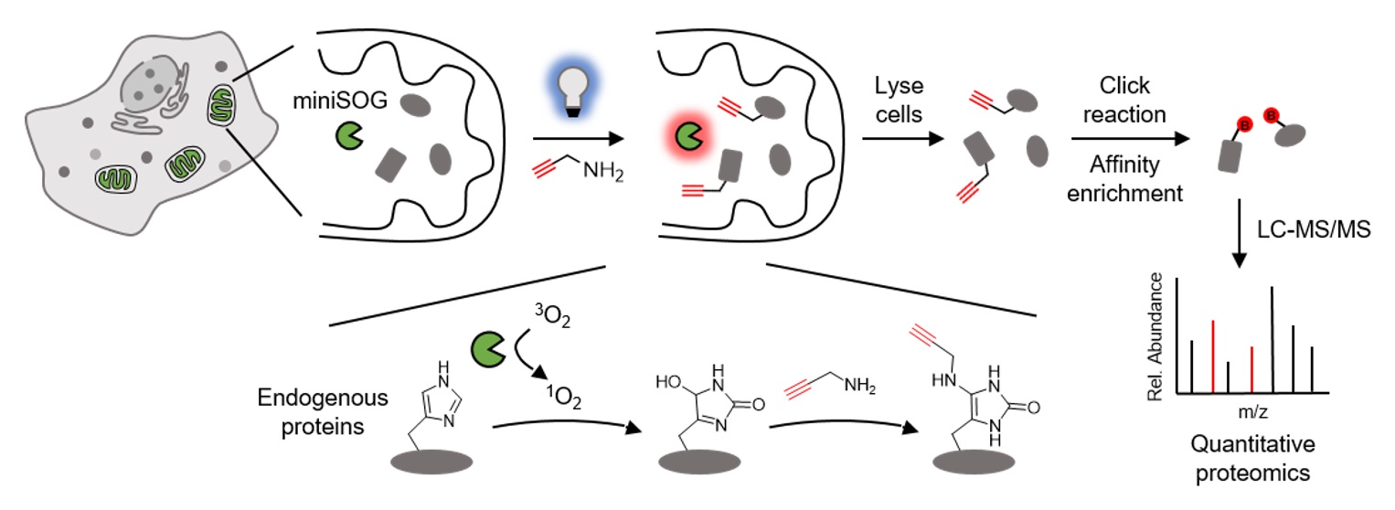
There has been a missing piece in our previous work on photocatalytic RNA labeling and DNA labeling with CAP-seq: in addition to nucleic acids, proteins too could get oxidized with photo-controlled release of singlet oxygen. Mapping the subcellular proteome with genetically encoded photocatalyst is appealing, as the light trigger offers precise temporal control of labeling. With patterned illumination schemes, it is even possible to spatially modulate labeling. Published in Nature Communications, we report the development of photocatalytic protein labeling with a genetically encoded protein tag, miniSOG. Our method, called RinID, can be broadly applied in multiple organelles, including the mitochondrial matrix, nucleus and the endoplasmic reticulum (ER). The temporal control of RinID enables pulse-chase labeling of ER proteome in HeLa cells, which reveals substantially higher clearance rate for secreted proteins than ER resident proteins. Now the toolbox is complete! Also check out our recent review of the state-of-the-art in the field of photocatalytic proximity labeling for profiling the subcellular organization of biomolecules, published in Chembiochem.
-
2023-03-30 Mar
Lab Stars!
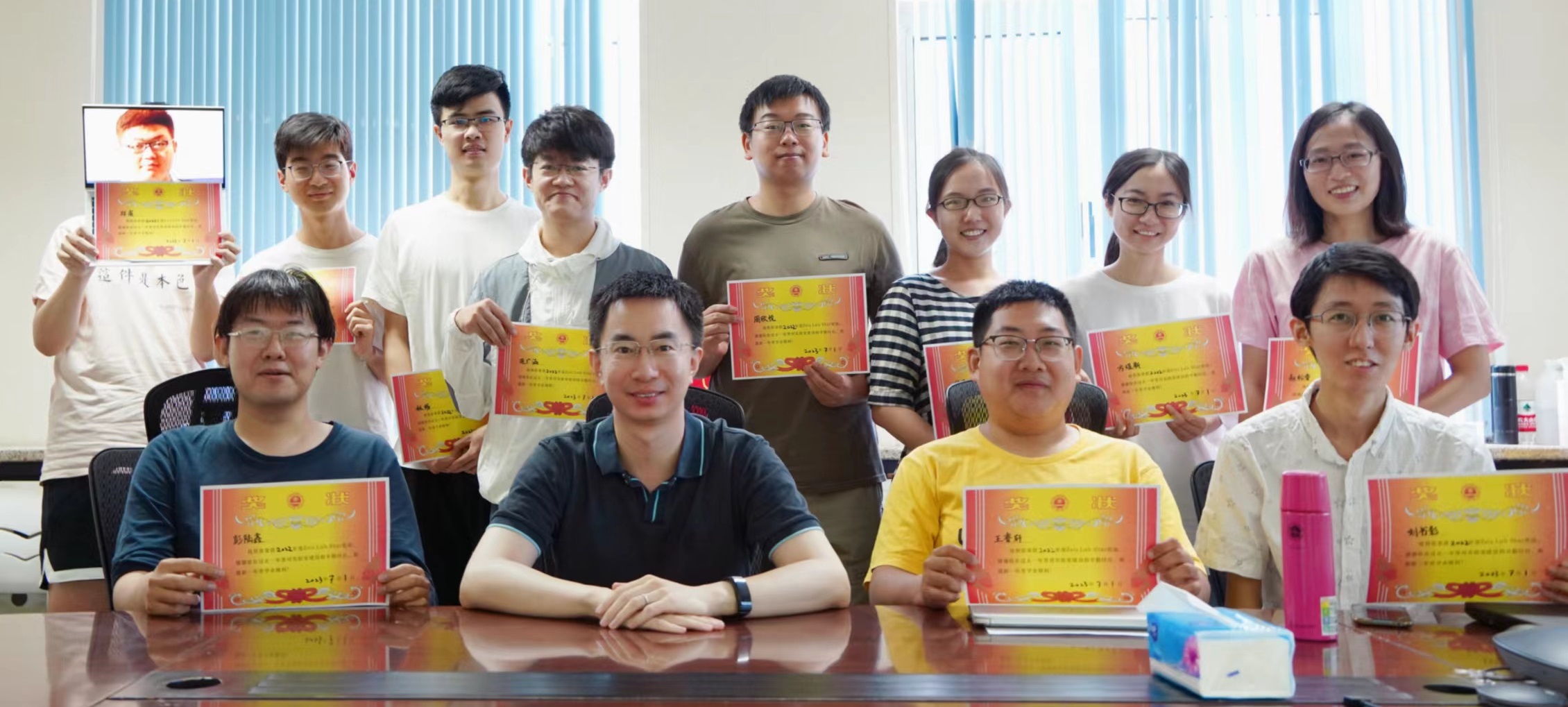
The Lab Star 2022 awards are presented to: (front row, from left to right) Luxin Peng, Ruixiang Wang, Shuzhang Liu, (back row, from left to right) Fu Zheng, Yangluorong Liu, Chang Lin, Guanghan Yuan, Xinyue Zhou, Ziqi Ren, Yuxin Fang, and Songrui Zhao. Thank you all for your dedicated contributions to the lab!
-
2023-02-15 Feb
Bioconjugate Chemistry Editor
Peng joins the editorial team of the ACS journal Bioconjugate Chemistry as a Topic Editor. Check out the February issue introducing the editorial team members. Under the leadership of our Editor-in-Chief, Prof. Theresa Reineke, we look forward to promoting the journal among young investigators and in the chemical biology community.
-
2023-01-12 Jan
Happy 2023!
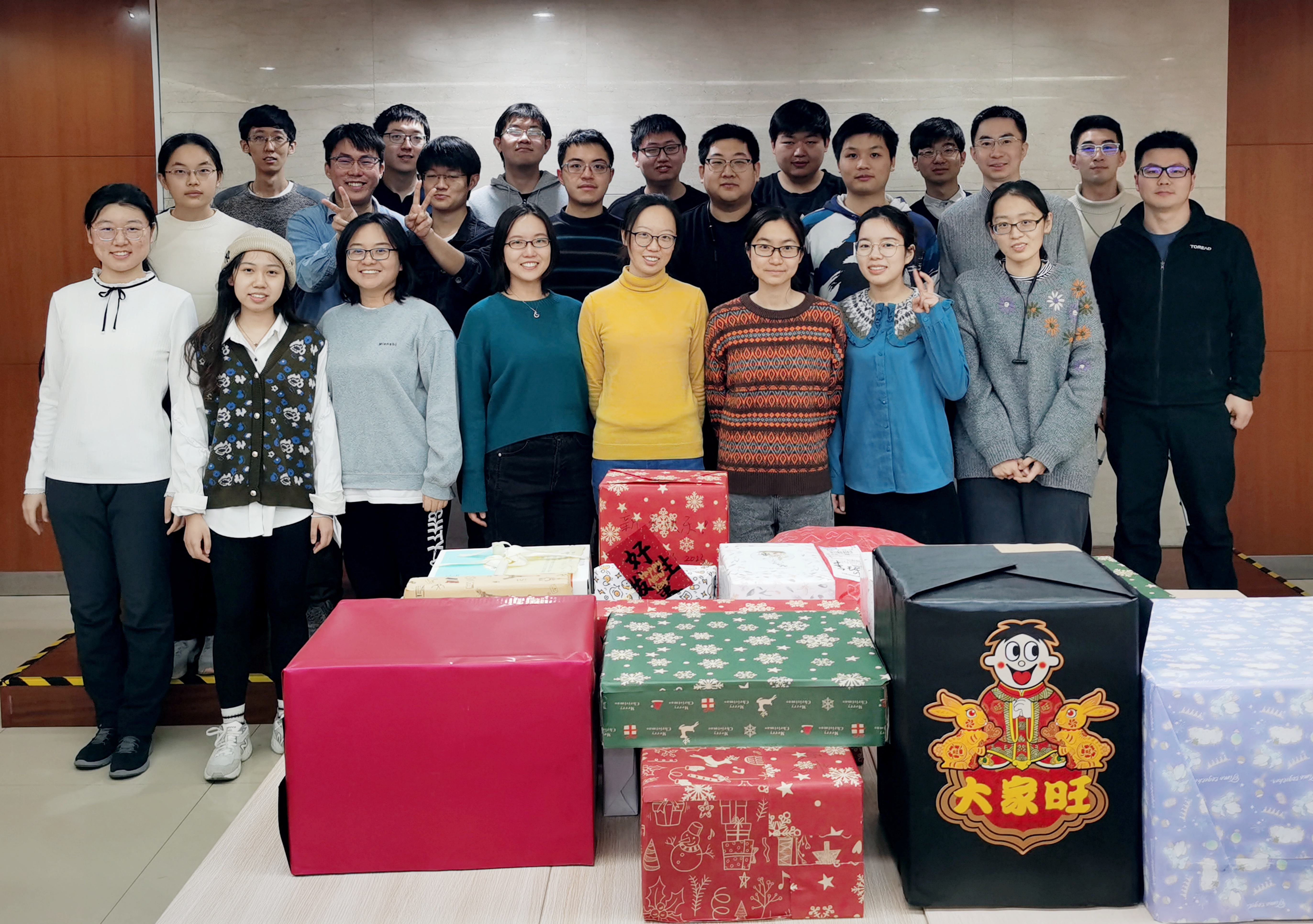
2022 has been a tough year. But looking at the photo, we have all survived and thrived. Our annual gift exchange continued on, and we are looking forward to a healthy and happy post-COVID 2023!
-
2022-08-19 Aug
Watching Action Potentials in Action

Voltage imaging offers an unique opportunity to measure the dynamics of action potential initiation and propagation, with a sub-micrometer spatial resolution that is unattainable by conventional patch-clamp electrophysiology. In two recently published research articles, we collaborated with Profs. Yan Zhang and Louis Tao at PKU to image action potential dynamics in cultured primary neurons with rhodopsin-based GEVIs. We mapped the velocity of action potentials at the axon initial segment (Zoological Research) and followed its back-propagation into somatodendrites (Neuroscience Bulletin). The observed action potential dynamics are explained through computational simulation.
-
2022-06-28 June
Farewell, Graduates!
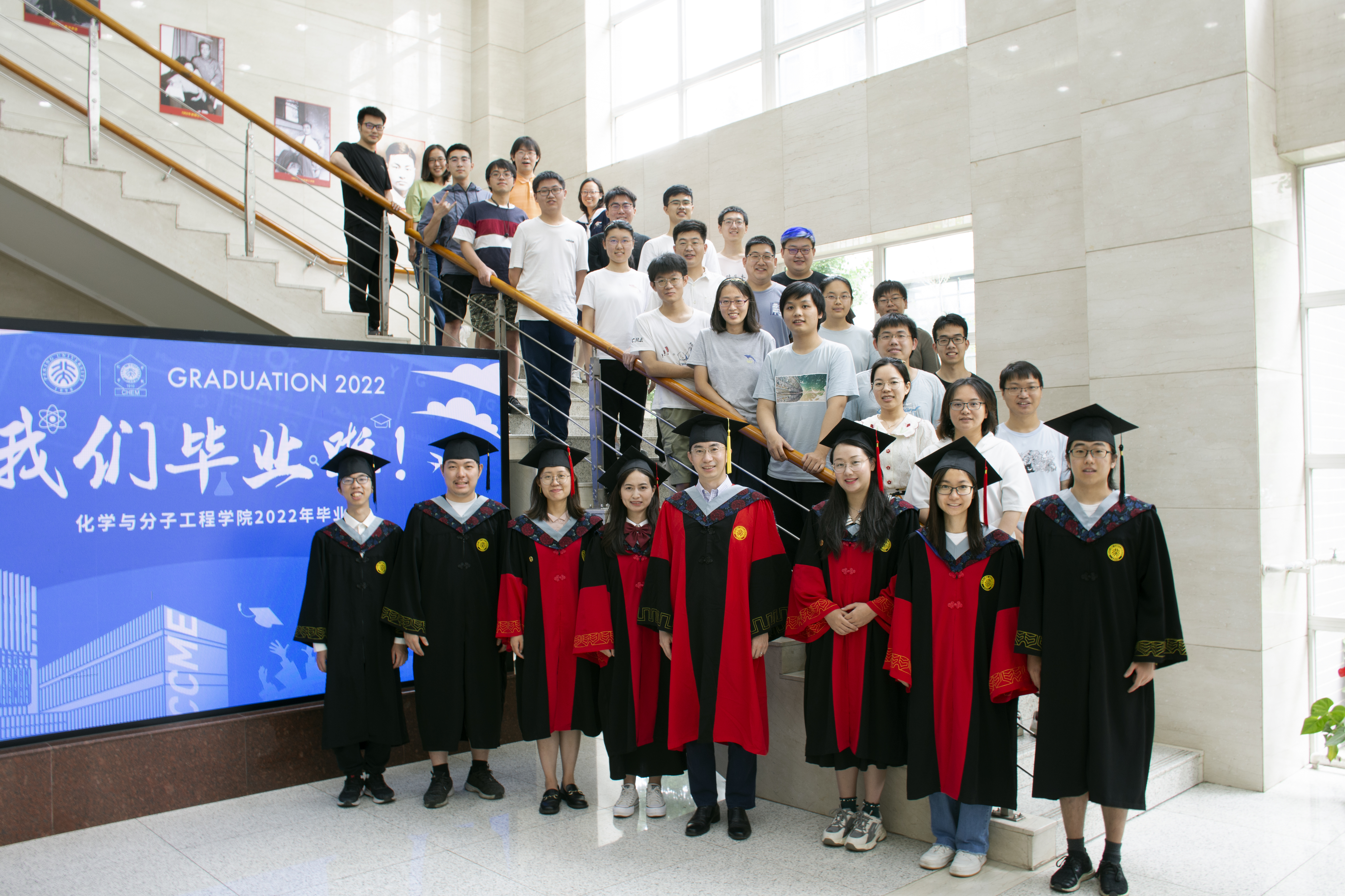
Congratulations to Yuxin, Ruirui, Feng, and Ran for defending their doctoral thesis! Yuixn will stay on as a Postdoctoral Fellow; Feng will be teaching biology at the High School Affiliated to SUSTECH in Shenzhen; Ran decides to join Sironax as a Research Scientist; Ruirui will work in the public sector in Chengdu. Wish you all great success in your chosen career paths! We are also proud of our three senior undergraduates (Yi, Lihao, Xuefeng) for receiving their BS degrees!
-
2022-05-05 May
Photocatalytic Crosslinking

The dynamic RNA-protein interactions play crucial roles in cellular physiology, yet characterizing these interactions in their native cellular context at the proteomic level has remained challenging. Published in Angewandte Chemie International Edition, we report a photocatalytic crosslinking (PhotoCAX) strategy for studying the composition and dynamics of protein–RNA interactions via mass spectrometry (PhotoCAX-MS) as well as RNA sequencing (PhotoCAX-seq). By integrating the blue light-triggered photocatalyst with a dual-functional RNA–protein crosslinker (RP-linker) and the phase separation-based enrichment strategy, PhotoCAX-MS reveals the dynamic change of RBPome in macrophage cells upon LPS-stimulation, as well as RBPs interacting directly with the 5′ untranslated regions of SARS-CoV-2 RNA. This work is the result of a collaboration with Prof. Peng Chen's group at PKU.
-
2022-03-03 March
Enhanced APEX-seq
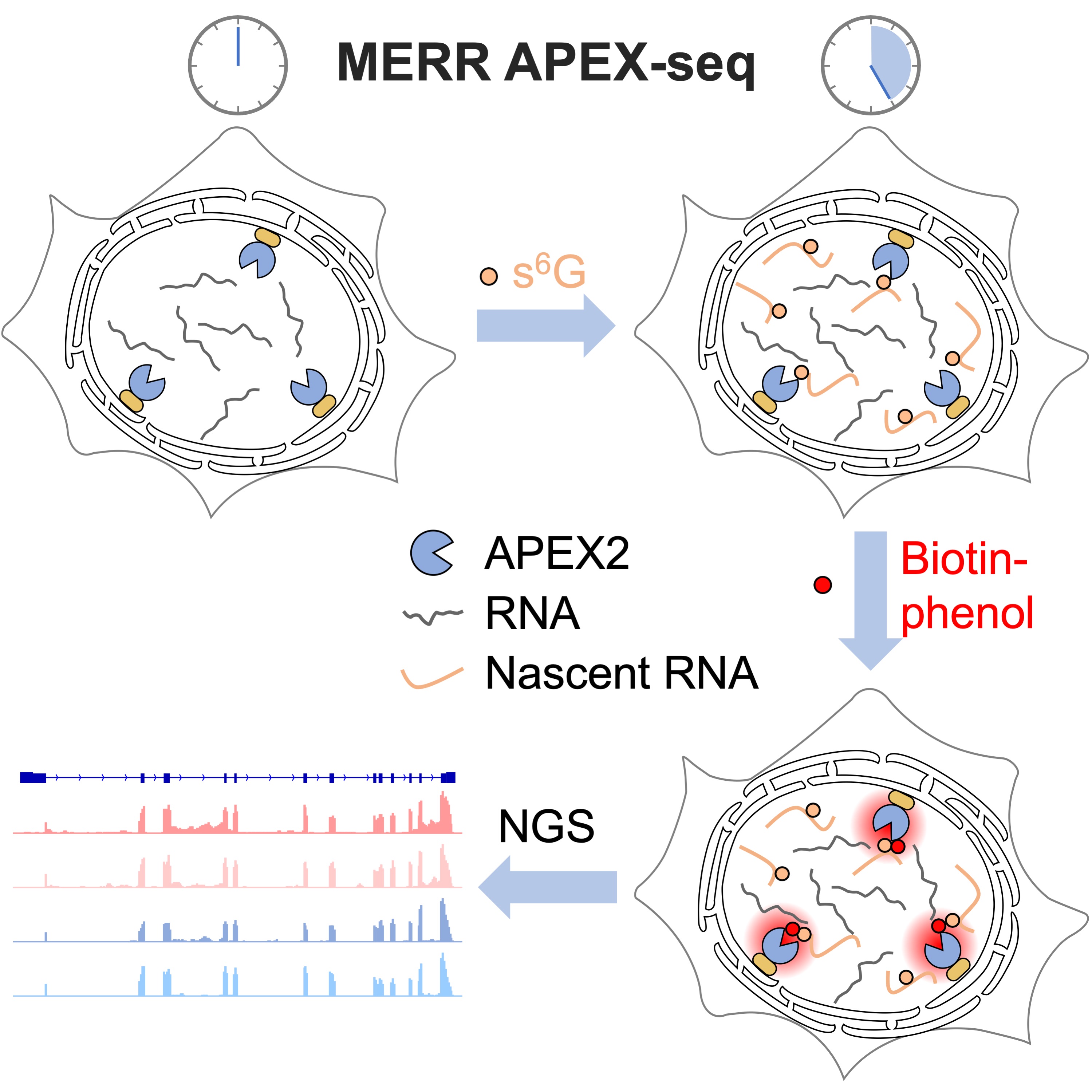
We have reported a sensitive method for profiling newly transcribed RNA with subcellular resolution in live cells. Published in Cell Chemical Biology, our method combines APEX-seq with non-canonical ribonucleoside metabolic incorporation, which enhances the biotinylation efficiency of APEX labeling. Application of this technique to the nuclear lamina reveals local transcripts involved in histone modification, chromosomal structure maintenance, RNA splicing and processing.
-
2022-02-20 Feb
Welcome Chenxin and Luming!
Chenxin Yu from Lanzhou University and Luming Yang from Xiamen University will complete their undergraduate thesis research in our lab. Both are incoming graduate students at CCME in the next semester. Welcome to join us!
-
2022-01-01 Jan
Happy 2022!
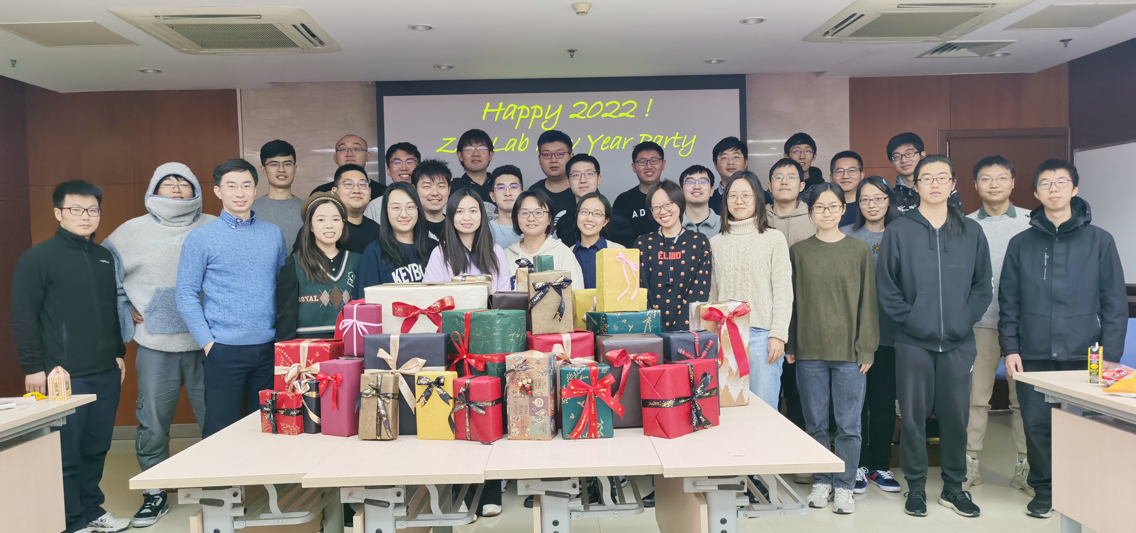
We celebrated New Year by exchanging gifts and sharing our loving memories of 2021. We look forward to a happy, healthy, and productive 2022! Our 2021 Lab Stars are Ruixuan, Shuzhang, Luxin, Yuxin, Feng, Ran, Gang, Wei, Chang, Ying, and Xinyue. Congratulations, and thank you all for helping make our lab a better place!
-
2021-09-15 Sept
Hybrid Indicator Review: Bringing Together the Best of Chemistry and Biology
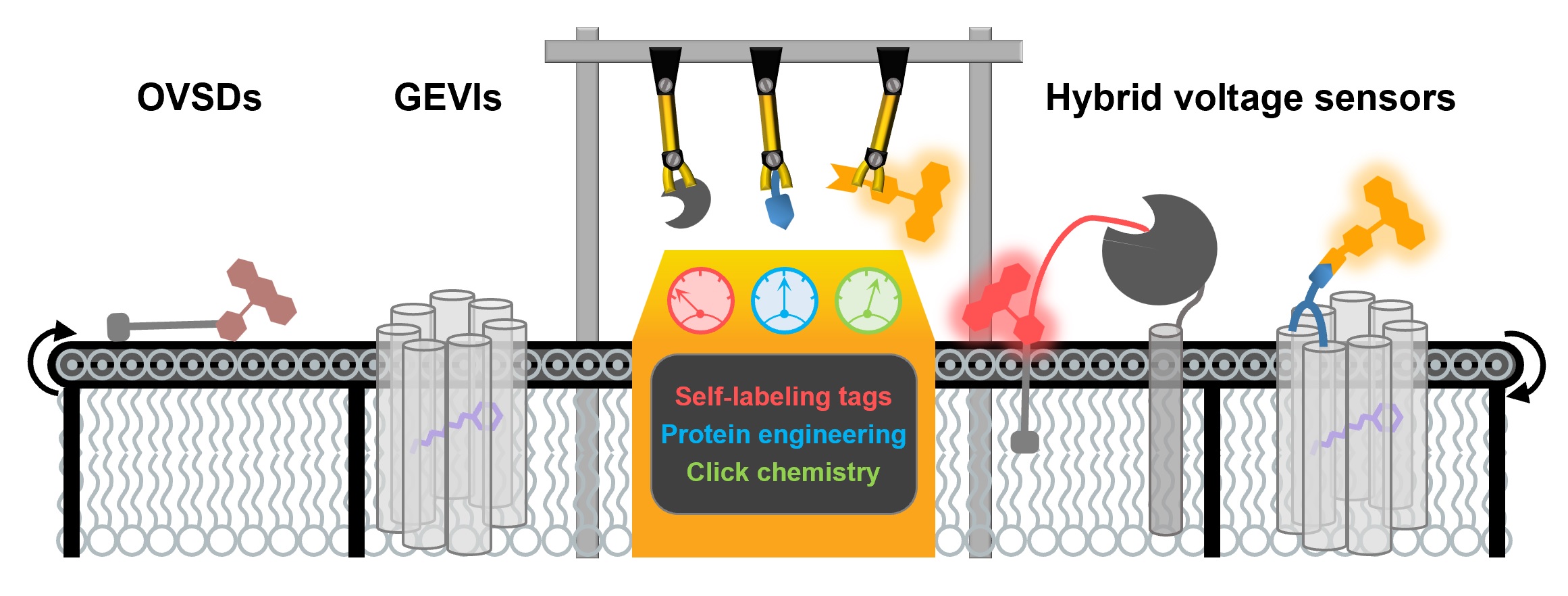
Check out our recent review article on Hybrid Indicators for Mapping Membrane Voltage, published in Journal of Neuroscience Methods! We summarize the state-of-the-art in the field of hybrid voltage indicators, including their molecular designs, cellular and in vivo characterizations and biological applications. We also discuss future improvements in biocompatibility, brightness and sensitivity of hybrid voltage sensors.
-
2021-08-12 Aug
Peng is Promoted to Tenured Associate Professor
The promotion recognizes our six years of efforts on developing chemical tools for studying neuroscience. Peng is grateful for all lab members, past and present, for their hard work. We are looking forward to launching the next phase of research at the interface of chemistry and neuroscience.
-
2021-06-27 June
Welcome, Undergrads!
Four students joined us this year for their undergraduate research training program. Welcome Wenyu Ding, Han Wang, Ruijie Li, and Peiyuan Meng!
-
2021-06-19 June
Congratulations to Our Graduates!

Over the past month, four graduate students (Wei, Ying, Tao, Anqi) and five undergrads (Luorong, Ziqin, Songrui, Guanghan, Zhiwei) have defended their theses. Congratulations to all of you! We celebrated these special occasions with a lab retreat that featured both stunning stalagmite and a fierce water fight.
-
2021-06-17 June
Creating the SubMAPP
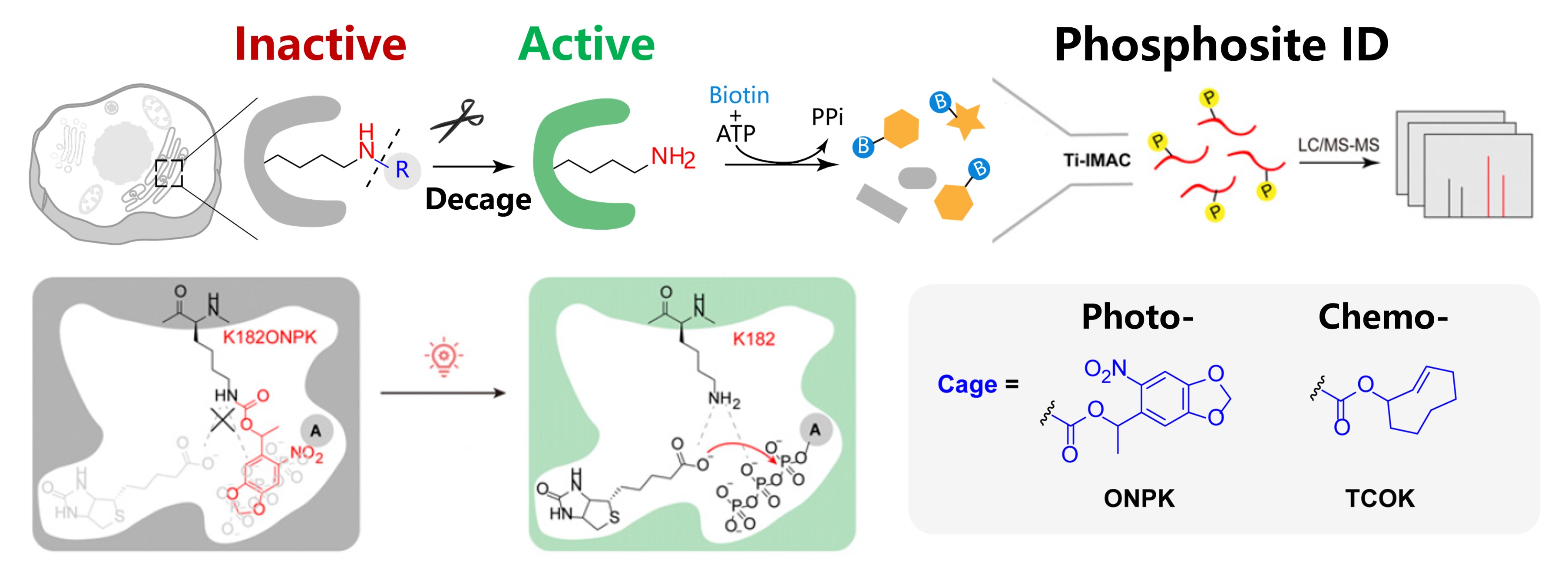
What happens when proximity labeling meets protein post-translational modification profiling? In collaboration with Prof. Peng Chen's group at PKU, we have developed a method for mapping subcellular phosphoproteome in living cells and animals. Our method, called SubMAPP, capitalizes on the genetically encoded bioorthogonal decaging strategy, which enables the rapid activation of subcellular localized proximity labeling biotin ligase through either light illumination or small-molecule triggers. This is then followed by the orthogonal pull-down of phosphopeptides and quantitative protein mass spectrometry analysis. SubMAPP allows for the investigation of subcellular phosphoproteome dynamics, revealing the altered phosphorylation patterns of endoplasmic reticulum luminal proteins when cells are under stress.
-
2021-05-04 May
Welcome Yu Chen and Chen Li!
Yu and Chen are enrolled in the CLS graduate program and have just finished their rotations. Both were undergraduates at CCME and trained in physical chemistry (Yu) and organic chemistry (Chen) labs. Welcome to join us and bring in new skill sets to our lab! They will work on projects related to spatial transcriptomics.
-
2021-04-17 April
Next-generation Hybrid Indicators
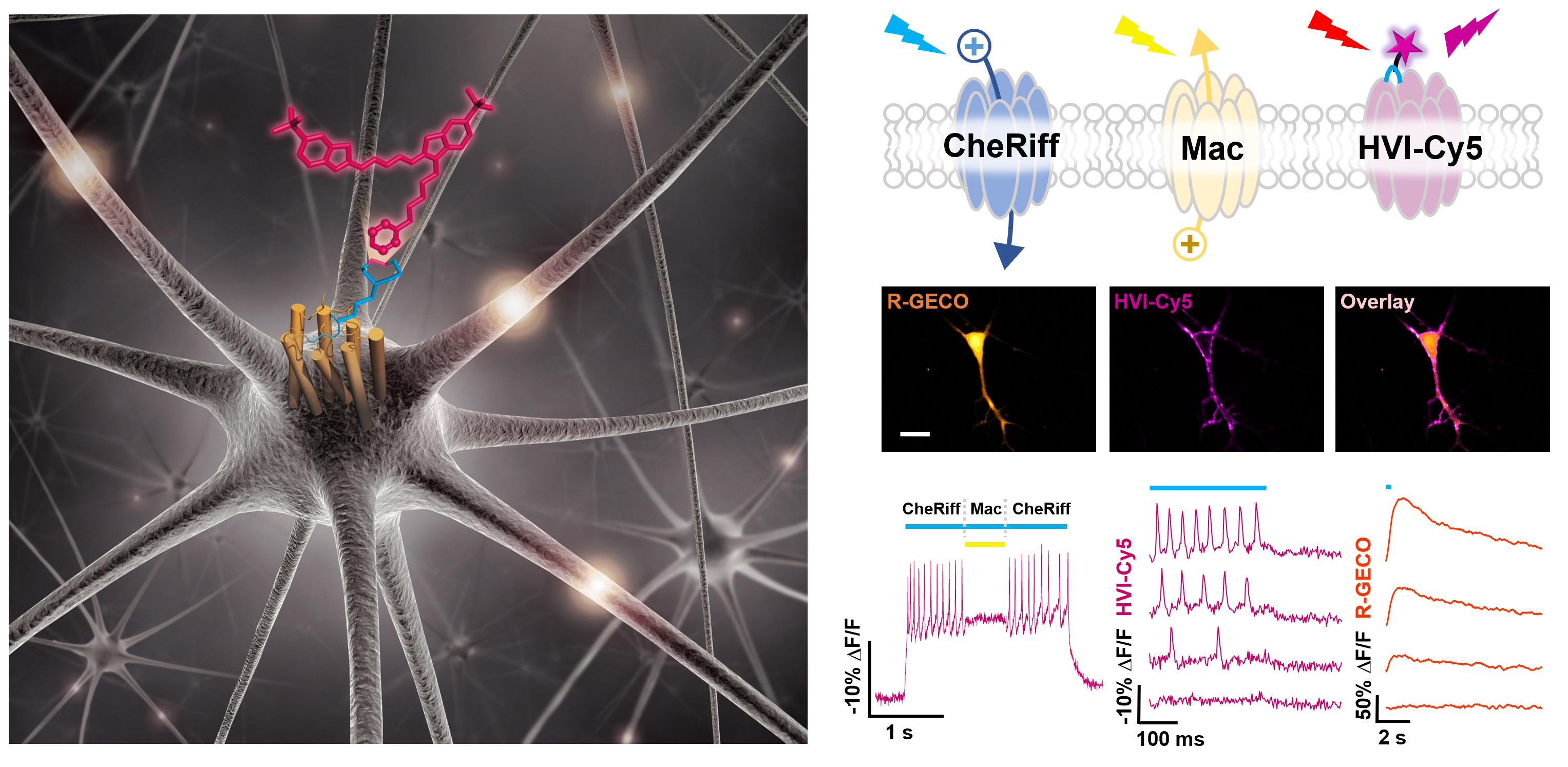
Our hybrid voltage indicator (HVI) paper is officially online at Nature Chemistry! Since the inception of Flare indicator in 2018, we have been working hard to implement this strategy in live neurons. Thanks to Diels-Alder reaction, we are able to assemble a palette of bright and sensitive HVIs in cultured rat hippocampal neurons. The most red-shifted HVI-Cy5 faithfully reports neuronal spikes in the far-red emission range and could be paired with optogenetic actuators and green/red-emitting fluorescent indicators, allowing multiplexed imaging and all-optical electrophysiology. We look forward to using this expanded toolbox in the high-throughput screening of neuronal ion channel agonists/antagonists. Congratulations to Shuzhang, Chang, Yongxian, and our collaborators in Prof. Peng Chen's group at PKU. To read more about the history of this work, check out our "Behind the Paper" post at Nature Portfolio Chemistry Community.
-
2021-01-01 Jan
Happy New Year!
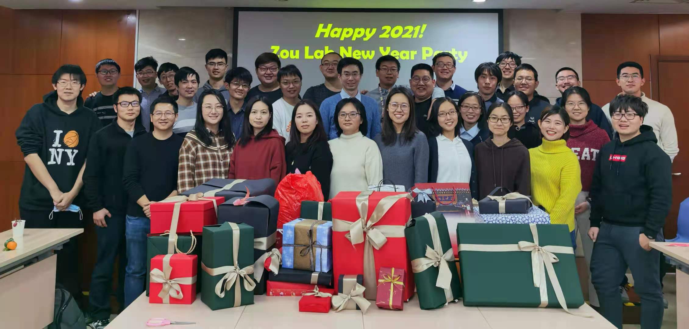
Although 2020 has been a difficult year for all of us, we are doing a great job coping with COVID-19. Surviving and thriving, we are looking forward to a brighter 2021! In our annual party, we exchanged gifts and shared memories of the past year. Congratulations to our 2020 Lab Stars - Shuzhang, Ruixuan, Luxin, Feng, Ran, Wei, Yi, Ying, Ruirui, Yuxin, Gang, Junqi, Ruixiang, and Chang. Thank you all for working hard to make our lab a better place!
-
2020-09-24 Sept
Bayer Investigator
Peng is honored to receive 2020 Bayer Investigator Award from Bayer-Peking University Center for Translational Research.
-
2020-08-17 Aug
CIBR Young Scholar
Peng is selected as CIBR Young Scholar by Chinese Institute for Brain Research (CIBR).
-
2020-07-09 July
Welcome Dr. Zhang!
We are delighted to welcome Dr. Wei Zhang to join our group as a postdoctoral fellow! Wei did his graduate study in molecular neuroscience at the Cajal Institute of Spanish National Research Council, where he investigated the mechanisms by which neurons establish and maintain cellular polarity. He obtained his PhD from Universidad Autónoma de Madrid (UAM) in Spain at the end of 2019.
-
2020-07-02 July
Cloud Commencement Ceremony
For the first time, we are having the Commencent ceremony online. Congratulations to Chaoling, Fu, Tenghui, and Wentao for successfully defending their undergraduate thesis and obtaining Bachlor of Science degrees! Chaoling, Fu, and Wentao will continue their PhD study in our department, while Tenghui has decided to defer his enrollment at the Scripps Research Institute at Florida Campus for one year to stay with us as a research assistant. Wish you all continued success!
-
2020-07-01 July
Welcome, Undergrads!
We have an unusually large pool of students joining our lab for the Undergraduate Research Training Program this year. Welcome Haoyun An, Rui Fu, Yi Han, Haoran Hu, Wenyu Kuang, Lihao Liu, Xiao Niu, Xuefeng Wang, and Zekun Wang! They will work on a diverse array of topics, ranging from super-resolution microscopy to directed evolution.
-
2020-05-29 May
APEX Goes Microbial!
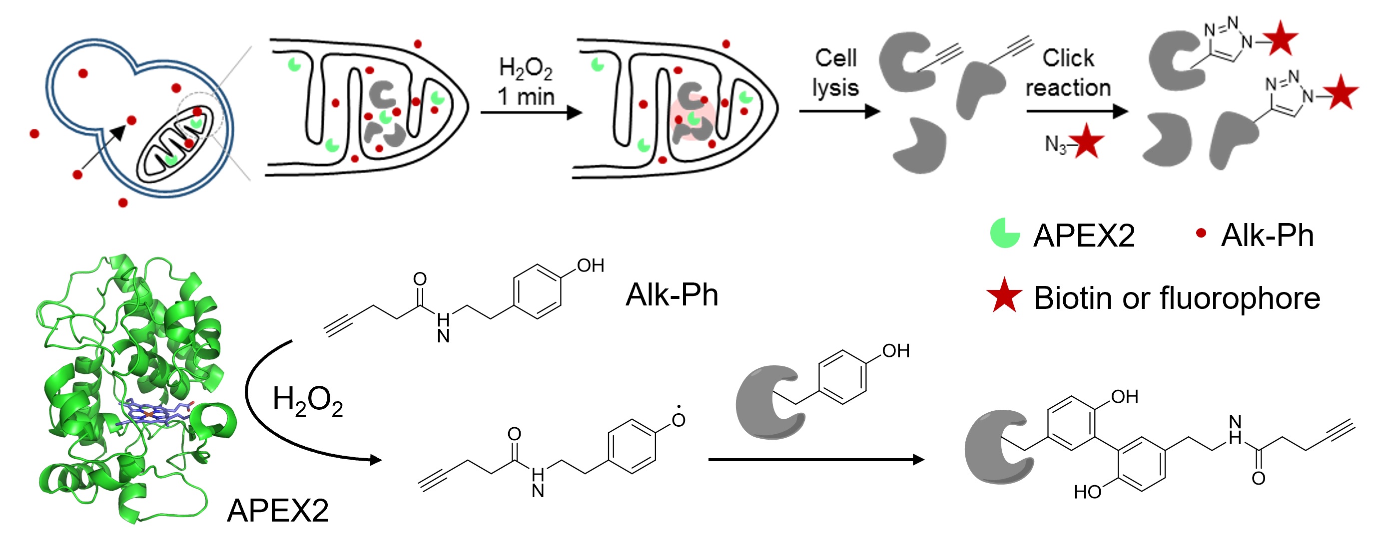
Check out our recent development of APEX labeling in yeast at Cell Chem Biol! By using a clickable APEX2 substrate, Alk-Ph, we have achieved proximity-dependent protein and RNA labeling in intact yeast cells. This new approach enables the proteomic and transcriptomic mapping of yeast mitochondrial matrix with high spatial resolution and coverage, thus extending APEX applications to microorganisms. We thank our collaborator, Dr. Jing Yang from Beijing Proteome Research Center. Congratulations to Yi!
-
2020-05-15 May
CAPA Award
Peng is honored and humbled to receive the 2020 OKeanos-CAPA Young Investigator Award at the Chemical and Biology interface. We thank CAPA (the Chinese-American Chemistry and Chemical Biology Professors Association) for the recognition of our work in chemical neuroscience.
-
2020-03-30 March
New Graduate Students
Chang Lin and Ziqi Ren officially joined our lab as first year CCME graduate students! Both of them did undergrad research here, working on fluorescence voltage indicator and proximity-dependent RNA labeling. Welcome both of you!
-
2020-02-18 February
Meeting Online
We resumed group meetings amidst the coronavirus outbreak, but had to use an online forum. Stay healthy, stay connected!
-
2020-01-14 January
A DNA trap
Congratulations to Yuxin's enTRAP-seq paper, published in the special issue of Biochemistry! As you read this news, your DNA is continuously under attack by reactive metabolites (particularly endogenous oxidants). Meanwhile, your cells are busy clearing up the resulting DNA damages, which have ramifications in tumorigenesis and gene expression control. To help illuminate the cellular mechanisms and the functional roles of oxidative DNA damage, we developed a genome-wide 8-oxoguanine profiling technique, termed enzyme-mediated trapping and affinity precipitation of damaged DNA and sequencing, or enTRAP-seq for short. When applied to analyze guanine oxidation in mouse embryonic fibroblast cells, enTRAP-seq revealed the enrichment of 8-oxoguanine in transcriptionally active chromatin regions.
-
2019-12-28 December
Happy New Year!
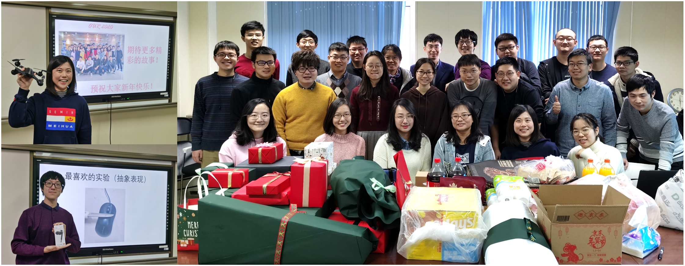
We exchanged gifts and memorable moments of 2019 at the New Year party. This year's Lab Stars are: Ruixuan, Luxin, Ran, Ying, Wei, Ruirui, Shuzhang, Gang, Yuxin, and Feng. Congratulations and thank you all for your hard work!
-
2019-10-22 October
SFBC Symposium Poster Award
Congratulations to Shuzhang for winning the poster award! He presented a bright and sensitive far-red voltage indicator for visualizing membrane potential dynamics in cultured neurons.
-
2019-10-09 October
An RNA Map
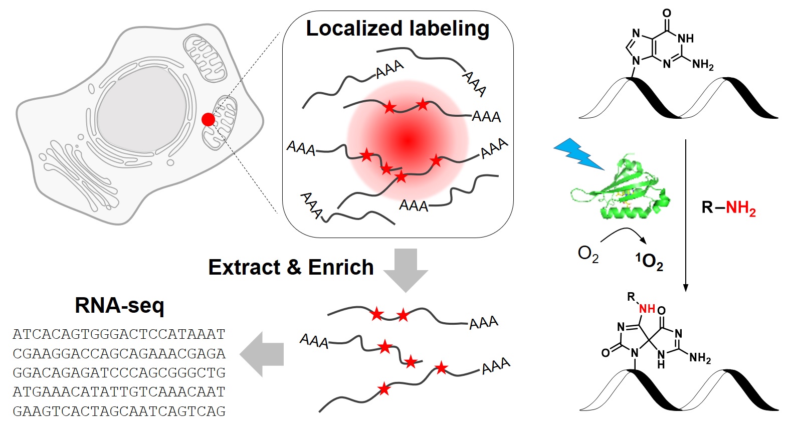
Check out our CAP-seq technique for mapping subcellular RNA localization at Nat Chem Biol! We employed a genetically encoded photosensitizer to achieve light-activated, proximity-dependent RNA oxidation, which allowed us to examine the local transcriptomes at various subcellular compartments. CAP-seq has high spatiotemporal resolution and is easy to use. We look forward to seeing more of its applications in the near future! We thank our collaborator, Prof. Jianbin Wang from Tsinghua University. Congratulations to Pengchong, Wei and Zeyao!
-
2019-09-10 September
Doctor Doctor!
Congratulations to Yongxian Xu (on 9/6) and Pengchong Wang (on 9/10) for defending their doctoral theses!
-
2019-08-27 August
Peng Selected as One of C&EN's Talented 12!
Peng Zou has been selected by Chemical and Engineering News (C&EN) as a member of this year's Talented 12 list. This program is presented annually by the newsmagazine and recognizes 12 young stars in the chemical sciences who are "working to solve some of the world's most challenging problems". This news was announced at the American Chemical Society National Meeting in San Diego, CA.
-
2019-07-04 July
Congratulations, Class of 2019 Undergrads!
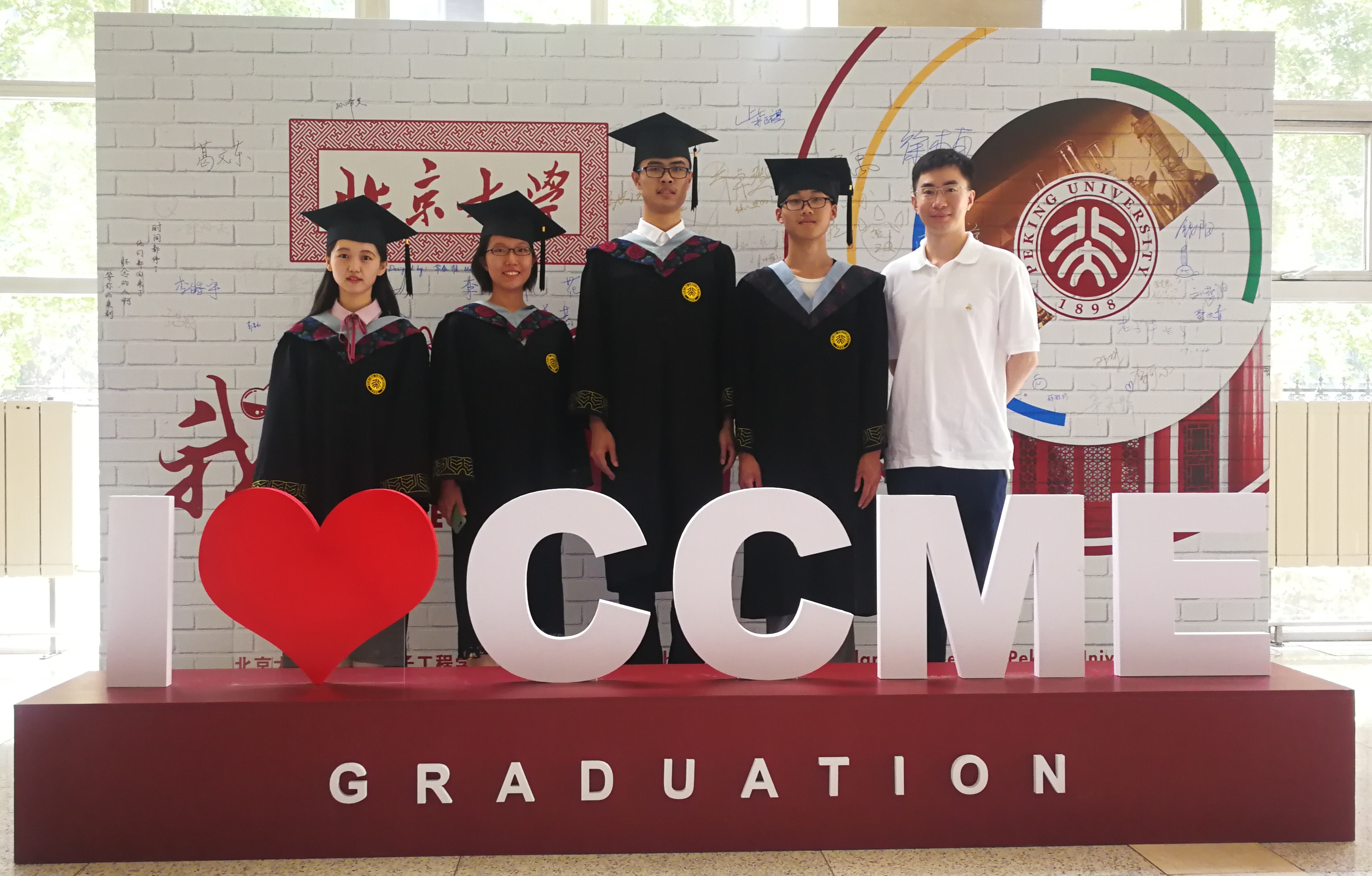
Congratulations to Xiaotian Bi, Ziqi Ren, Chang Lin, and Liyuan Zhu (left to right) for getting their Bachelor's degrees! Ziqi and Chang will continue their study at PKU as graduate students. Xiaotian (Caltech) and Liyuan (Princeton) will start their PhD journey in the US after the summer. We look forward to seeing your continued success in the years to come. Congratulations again to all of you!
-
2019-06-27 June
Pick Your Favorite Substrate

Check out our new panel of APEX substrates in Angewandte Chemie International Edition! We synthesized a series of aromatic compounds and identified arylamines as efficient probes for labeling RNA and DNA. Using the mitochondria as a model, we demonstrated proximity-dependent RNA capture with exceptional spatial-specificity and high coverage. Congratulations to Ying and Gang! This is our first venture into the RNA world.
-
2019-04-20 April
Welcome Yi, Ruixiang, and Xinyue!
Yi Li, Ruixiang Wang, and Xinyue Zhou joined our lab as first graduate students. Yi did his undergrad research in our group and has since decided to stay. Both Ruixiang and Xinyue were from Nankai University. They are enrolled in CLS program and have recently finished rotations. They will work on research projects related to proximity proteomic labeling.
-
2019-01-26 January
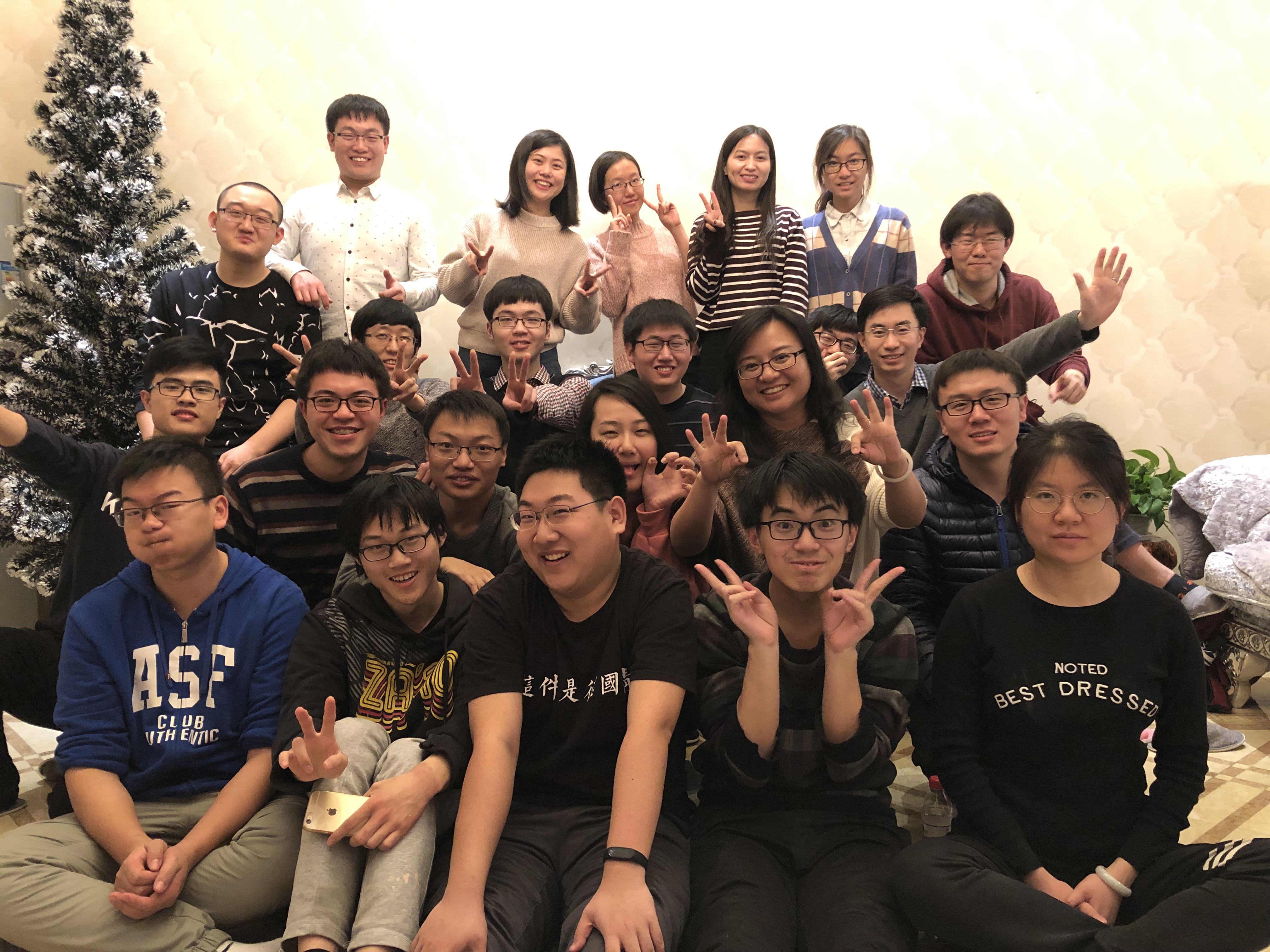
Happy Chinese New Year!
-
2018-12-28 December
Happy New Year!
As a lab tradition, we played gift swap games at the New Year party and shared our memory of 2018. This year's Lab Stars are: Luxin, Pengchong, Ruixuan, Shuzhang, Yongxian, and Wei. Congratulations to everyone!
-
2018-08-22 August
Poster Award, Again!
They did it again! Congratulations to Wei Tang and Pengchong Wang for winning Poster Award at the 5th Asian Chemical Biology Conference in Xi'an! We look forward to seeing broader applications of our technique in the years to come.
-
2018-07-10 July
Congratulations, Undergrads!
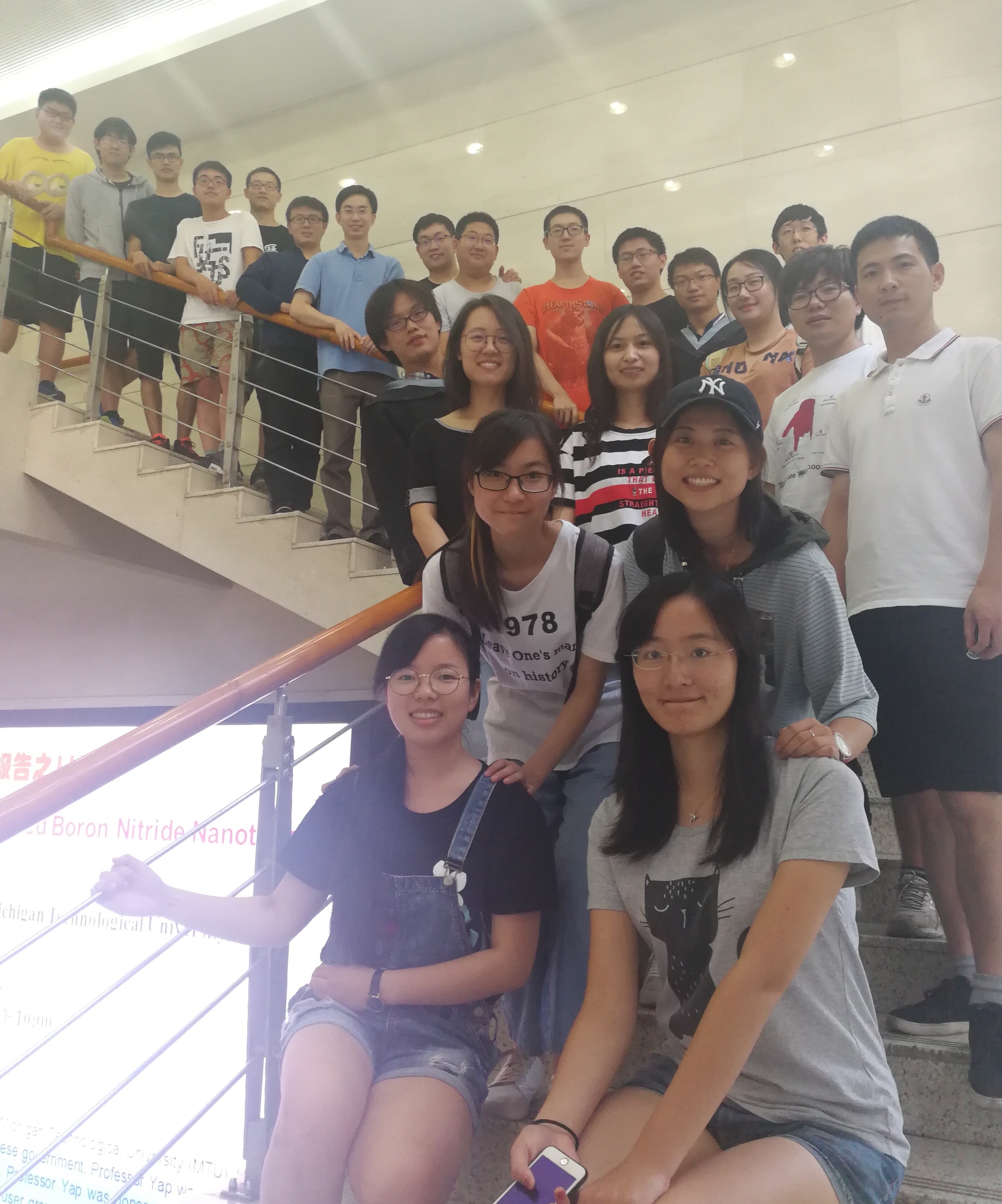
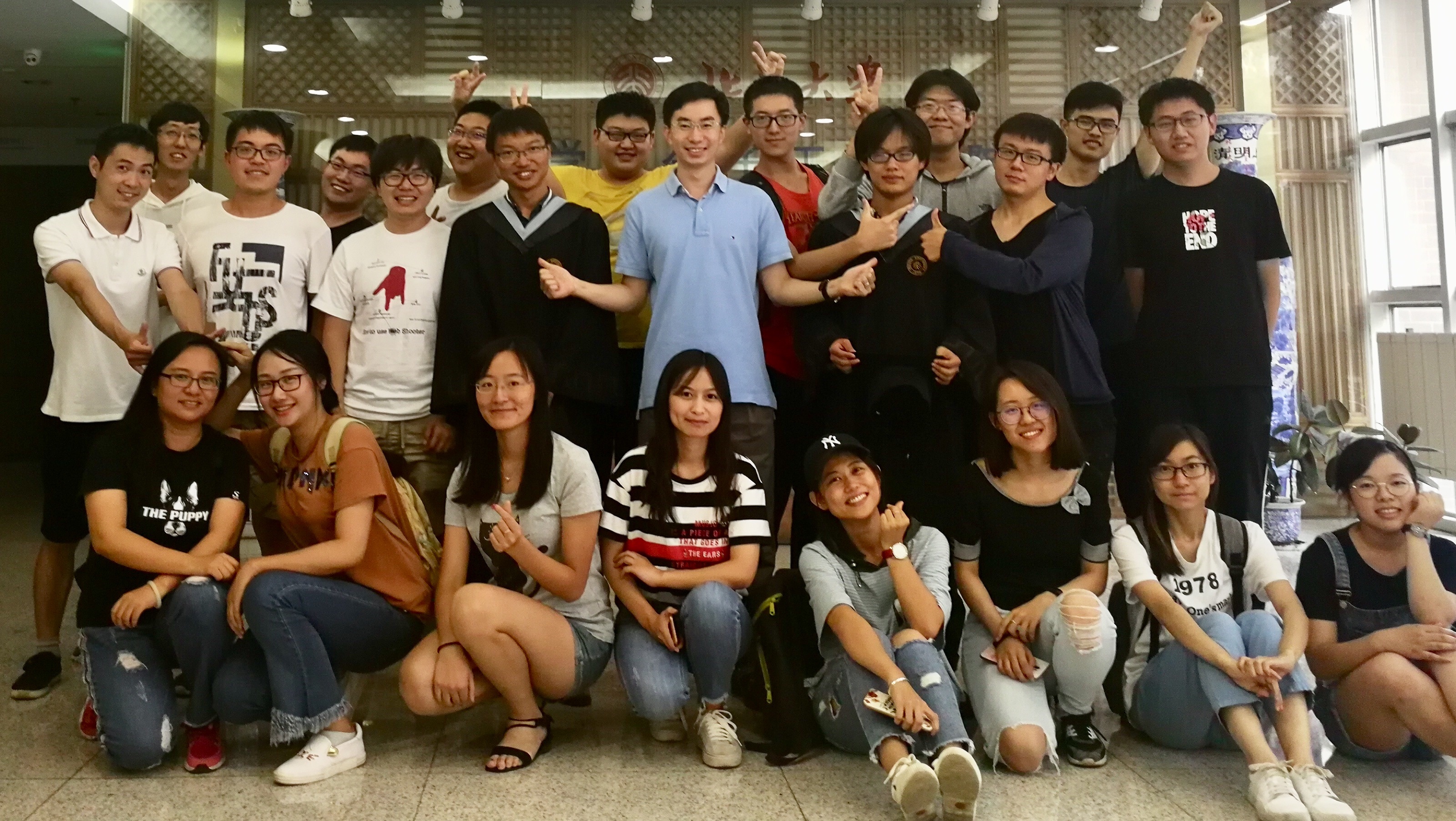
Congratulations to Yi Li and Zhongyu Zou for successfully defending their undergraduate theses! More Bachelor's degrees in Zou Lab! Zhongyu will soon join the Department of Chemistry at the University of Chicago as a graduate student. Yi will continue his PhD study in our department at PKU.
-
2018-05-18 May
Best Poster Award!
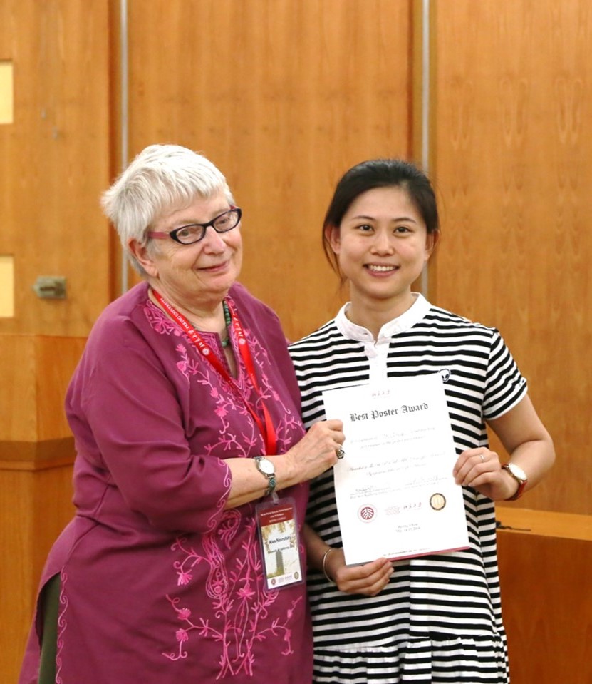
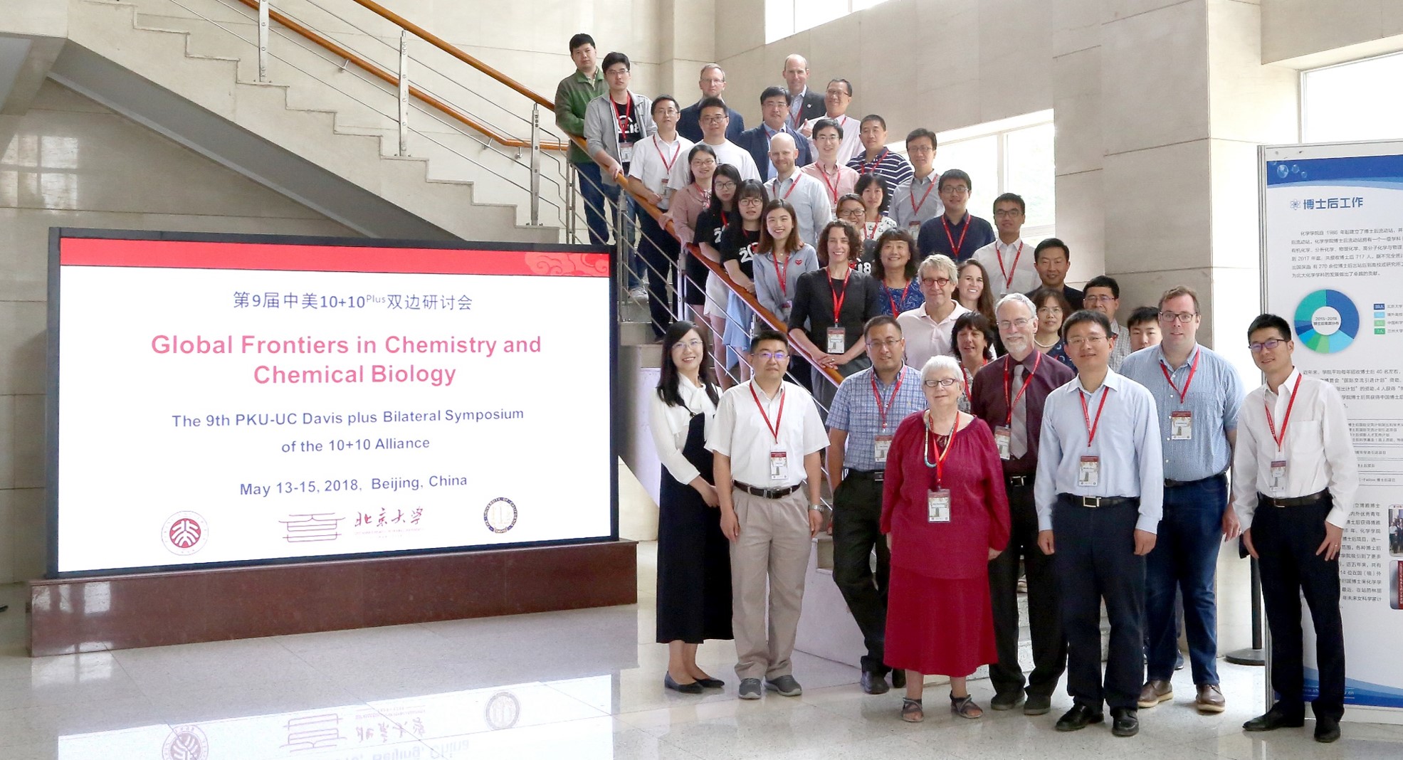
Wei Tang won the Best Poster Award at the PKU-UC Davis Symposium this week! During the symposium, each student gave a 3-min elevator pitch to highlight their research to the audience. Professor Alexandra Navrotsky from UC Davis presented Wei the award. The project, titled "Spatial specific RNA profiling via chromophore-assisted proximity tagging (CAP-tag)", was led jointly by Wei Tang and Pengchong Wang. Congratulations to both of you!
-
2018-04-21 April
New Students
Three graduate students joined our lab this month! Ran Li and Gang Wang are both Chemistry majors from the CLS program, while Junqi Yang is our first PTN student. Welcome all of you!
-
2018-03-06 March
Hybrid is Better!
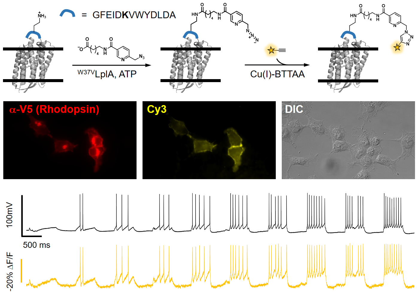
Our work on hybrid voltage indicator is accepted in Angewandte Chemie International Edition! By site-specifically modifying microbial rhodopsin on live cell surface with organic fluorophores, we created a palette of hybrid indicators with combined merits of small molecules (bright, photostable and small in size) and proteins (genetically targetable). The best of this series, called Flare1, has submillisecond response speed and superior sensitivity than the previous generation of fluorescent protein-based sensors. It allowed us to investigate the spatial and temporal correlation of electrical coupling among mammalian cells. Future developments are underway to achieve voltage imaging in neurons or in vivo. Read more about the design and deployment of hybrid indicators to neuroscience research in our recent review in ACS Chemical Neuroscience.
-
2018-01-01 January
New Year and New Students!
As 2018 approaches, we are delighted to welcome three CCME graduate students to join our group: Yuxin Fang, Shuzhang Liu, and Ruixuan Wang. Yuxin (B.S. in Chemistry from Nankai University) did her undergraduate thesis research in our group earlier this year and has since decided to stay. Shuzhang and Ruixuan graduated from USTC and PKU, respectively. At our New Year party this week, we exchanged gifts and shared our memorable moments in 2017. We also announced our annual Lab Stars award: Luxin, Pengchong, Ying, and Wei. Congratulations and thank you all for your hard work! We are all looking forward to a productive and happy New Year!
-
2017-06-26 June
Congratulations to Our Undergraduates!
Congratulations to Yu Wang and three Yuxin's (~ Fang, ~ Mi, ~ Duan) for successfully defending their theses and getting Bachelor's degrees! After the summer, they will all move on to the next stage of their academic careers. Yu Wang will join The Scripps Research Institute for her PhD study. Yuxin Fang will be enrolled in the PhD program at CCME, PKU. Both Yuxin Mi and Yuxin Duan will move to the UK to study at the University of Oxford.
-
2017-05-02 May
State-of-the-Art in Voltage Imaging
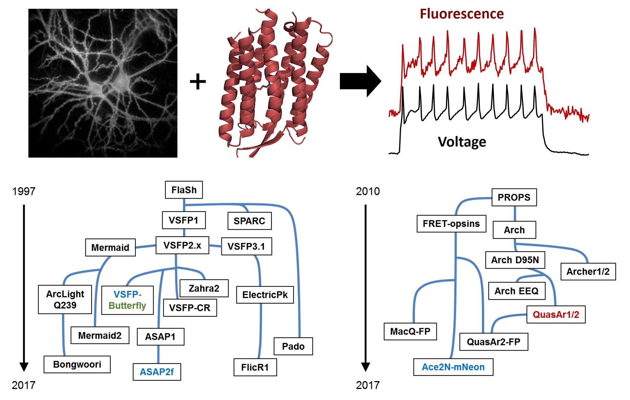
The past decade has seen a proliferation of fluorescent voltage indicators. Given the multiple choices, each with different tradeoffs between response speed and sensitivity, brightness and spectral range, one might ask: which sensor is better? In a recent review published by our group and Prof. Adam Cohen at Harvard University, we outlined the history of these voltage indicators, highlighting those with promising characteristics for in vivo imaging. We also discussed protein engineering strategies that gave rise to these sensors, and hoped that this article will inspire novel ideas for better probes. Read about it in Current Opinion in Chemical Biology.
-
2017-03-05 March
Welcome Ruirui and Yuxin!
We are delighted to have Ruirui Ma and Yuxin Fang joining our group. Ruirui is a first year CCME graduate student with prior research interest in inorganic and physical chemistry. We are excited to integrate that background into our research focus. Yuxin is currently a senior undergraduate student from College of Chemistry at Nankai University, and will be entering CCME in the coming fall term. Welcome both of you!
-
2017-01-03 January
New Students
As we enter the New Year, we are delighted to have four more students joining the group! Feng Yuan (B.S. in Biology), Anqi Wang (B.S. in Chemistry), and Tieqiang Yue (B.S. in Chemistry) all graduated from Cuiying Honors program at Lanzhou University. Luxin Peng majored in physical chemistry as an undergraduate at Nanjing University. This timely reinforcement will enable us to explore new research directions in the new year. Welcome all four of you!
-
2016-12-27 December
New Year Party and Lab Stars
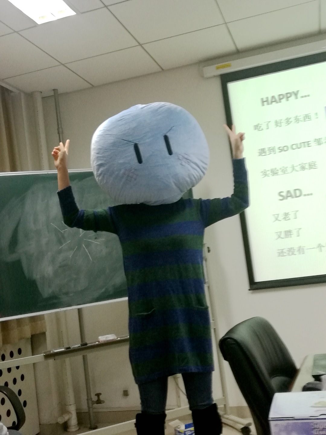
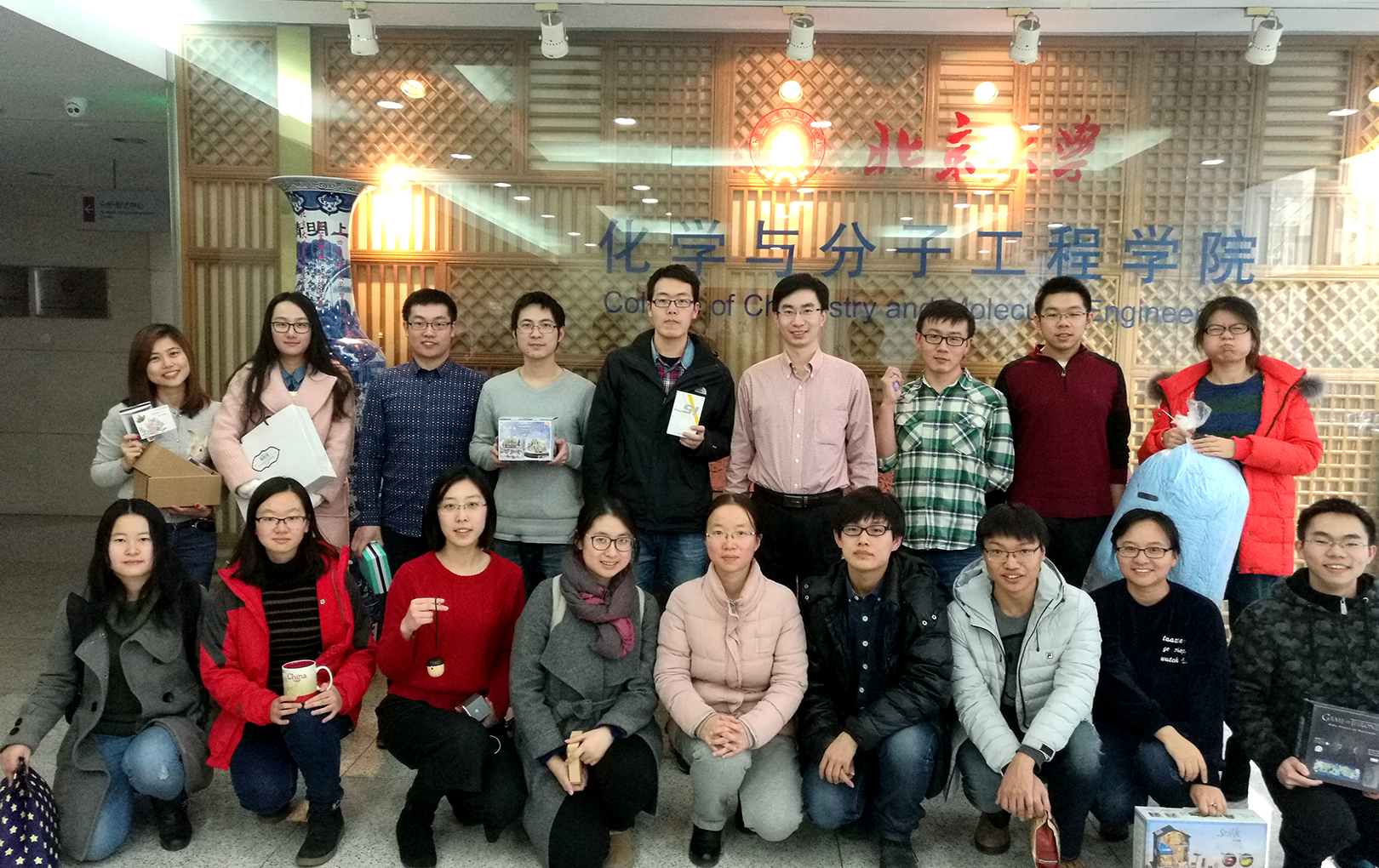
In addition to the gift swap game as a tradition, this year's party also included a one-slide flash talk, where each one of us reflected upon our joy and happiness and achievements in the past year. Old is gone and New is here. We are looking forward to a prosperous and productive 2017! As a new tradition, we have established our annual Lab Stars award, which will be determined at the end of each year by anonymous votes from a majority of lab members. This year, our first award goes to Sicong Wang, Ying Zhou, and Yongxian Xu, to recognize their strong work ethic and outstanding contributions to the lab.
-
2016-07-18 July
New Recruits and Summer Interns
Several students joined our group in the past couple of months. Wei Tang obtained her B.S. degree from Sun Yat-sen University and was enrolled in the CLS program at PKU in 2015. She started rotation in our lab this April and has decided to stay since. Sicong Wang joined us as a Research Assistant last month. He recently graduated from Beihang University (BUAA) with a Bachelor's Degree in Biomedical Engineering. We are also pleased to have Renjie Shang (Ocean University of China) and Yongpeng Jiang (Wuhan University) join us as summer intern students. Welcome all of you!
-
2016-07-06 July
Undergraduate Research Fellowship
Congratulations to Chenxi Sun (Yuanpei College, '18) and Shasha Feng (College of Life Sciences, '18) for being selected for Beijing Innovation Program for Undergraduate Research! Both awardees will receive research fund to support lab work. We are very proud of you!
-
2016-05-26 May
Thesis Defense
Congratulations to Jiahe for defending her undergraduate research thesis! Jiahe was the first undergrad to join our lab last summer, and has been working on developing the next generation voltage sensor for her thesis. Jiahe will move to UK this summer. She is the recipient of a graduate fellowship from China Scholarship Council, which will sponsor her PhD study at the University of Oxford for the next four years. Congratulations again!
-
2016-05-23 May
CNHUPO Young Investigator
Peng Zou is honored to receive the Young Investigator Award at the 9th CNHUPO (Chinese Human Proteome) Annual Congress (2016.5.21-23, Xiamen).
-
2016-01-09 January
Talent Award
Peng Zou has received Thousand Young Talents Award from Chinese Government. This will encourage and support future research efforts in our lab.
-
2015-11-11 November
More Rotation Students
Our lab size is temporarily doubled with the arrival of five rotation students! Jianzhi Zeng, Zhentao Zi and Shumei Hu are from CLS program. Hang Cheng and Chen Qi are from PTN program. They will start exploring new project ideas in the coming two months. Welcome!
-
2015-09-20 September
Rotation Students
Welcome Ying Zhou and Tao Ding, two CCME first-year grad students to start rotation in our lab. Ying obtained her Bachelor of Engineering degree from Soochow University. Tao graduated from CCME at Peking University.
-
2015-09-01 September
Undergrad for the New Semester
As the new semester starts, Jiahe Yang, a senior undergrad from School of Life Sciences, joins us to work on voltage sensor engineering for her thesis. Welcome on board!
-
2015-08-04 August
A Brand New Lab
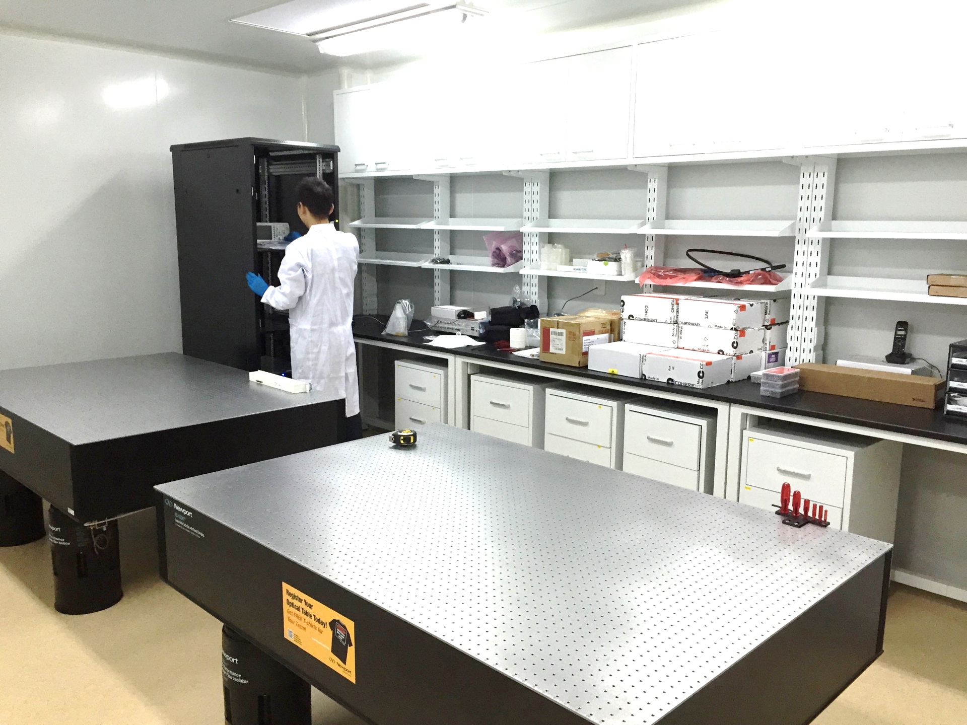
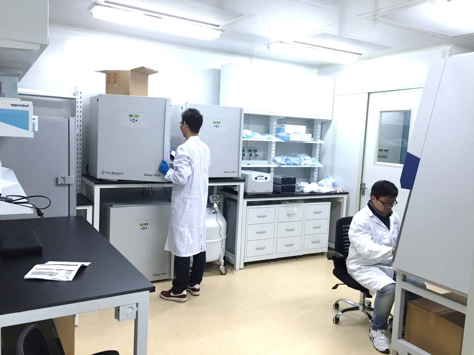
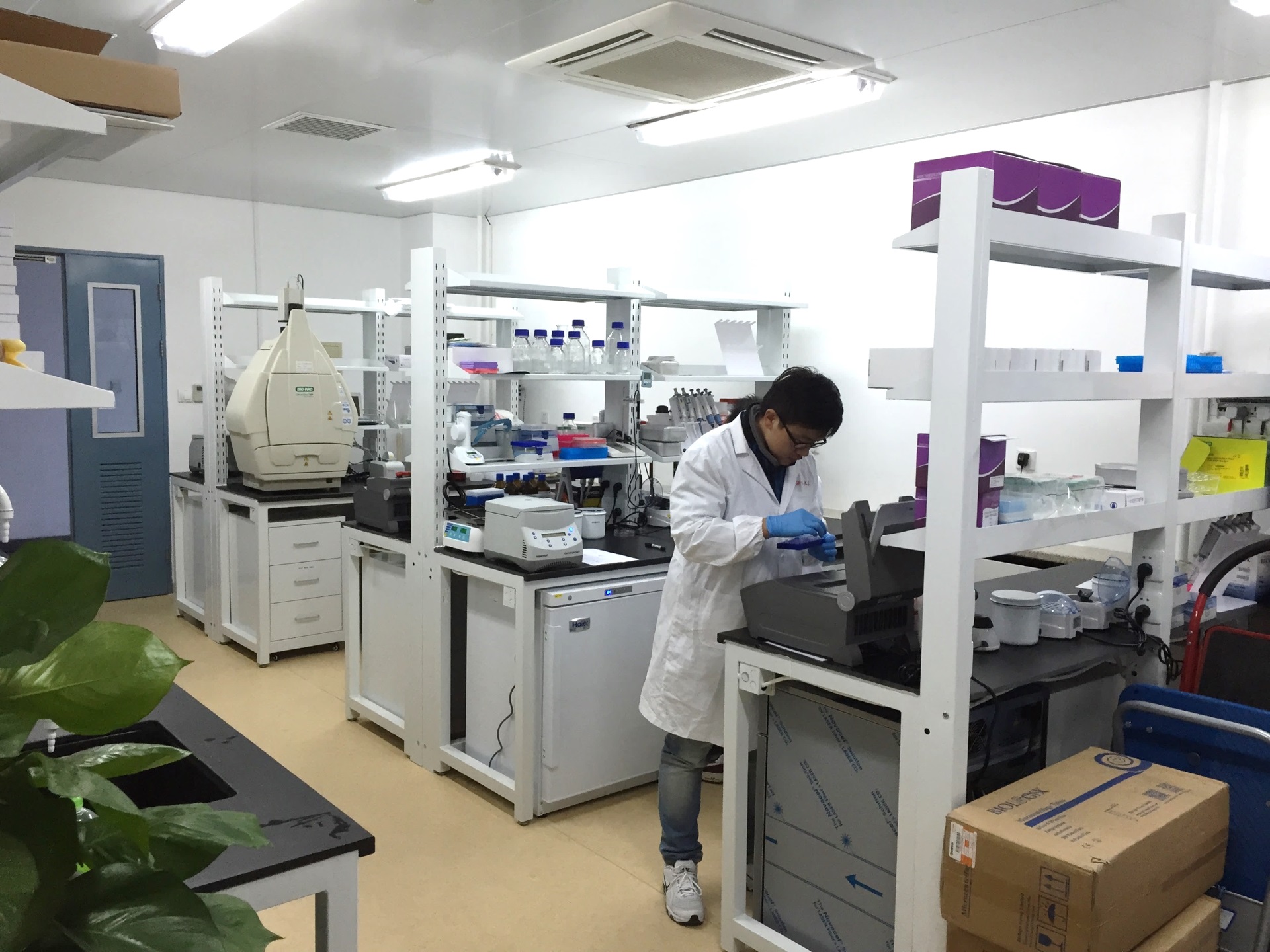
Lab renovation is completed. Our space is divided into four areas: optics room, tissue culture room, wet lab, and student office. The first two areas are built within a clean room.
-
2015-07-11 July
Lab Assistants
Tongyao Wei joins our lab as research assistant. He recently graduated from School of Life Sciences at Lanzhou University. Jing Zhang joins us as administrative assistant. Welcome both of you!
-
2015-06-15 June
We are Growing
Welcome the first two Zou lab members: Yongxian Xu and Pengchong Wang! Both are enrolled in the CLS program. Yongxian obtained his MS degree from Zhejiang University. Pengchong graduated from Shandong University.
-
2015-05-13 May
A New Chapter
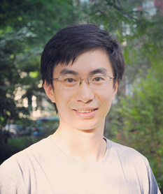
Peng Zou is appointed as an Assistant Professor in the College of Chemistry and Molecular Engineering at Peking University. He is also affiliated with the PKU-Tsinghua Center for Life Sciences, and PKU-IDG/McGovern Institute for Brain Research, as a Principal Investigator.


